Chapter 150
Pleomorphic Adenoma (Benign Mixed Tumor)
Epidemiology
Pleomorphic adenoma is the most common (80–90%) of all primary minor salivary gland tumors to arise within the parapharyngeal space. These tumors may occur in any age group, but they most often present between 40 and 50 years of age. Males and females are equally affected.
Clinical Findings
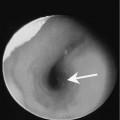
Patients usually present with a painless submucosal tonsillar mass. On physical examination, the mass is nonpulsatile and firm to palpation.
Pathology
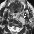
Parapharyngeal space pleomorphic adenomas are thought to arise from rests of minor salivary gland tissue located in the fat of the parapharyngeal space. On gross pathological examination, these tumors are encapsulated smooth round or oval masses. The terms pleomorphic and benign mixed refer to the histologic appearance of this neoplasm. The tumor consists of both epithelial and mesenchymal elements. The epithelial component consists of an inner layer of epithelial cells, whereas the outer layer consists of myoepithelial cells that constitute the mesenchymal component. Both elements must be present for the diagnosis of pleomorphic adenoma to be made. These tissue elements may be arranged in a variety of patterns and associated with a variable amount of stroma. The stroma can consist of variable amounts of mucoid, fibroid, chondroid, vascular, or myxochondroid elements. The mucoid stroma is thought to be responsible for the signal characteristics present on T2-weighted imaging.