Pleura


Pleura


Rama S. Ayyala

Shunsuke Nosaka

Khalid Khashoggi

Janina M. Patsch

Zaleha Abdul Manaf

Edward Y. Lee


INTRODUCTION

In children, various pathologic disorders can affect the pleura and the pleural space with imaging playing a vital role in determining the underlying etiology, location, and extension. Pleural abnormalities can be subtle and often require careful investigation with multiple imaging modalities in the pediatric population. This chapter provides an overview of the currently available imaging modalities to evaluate pleural abnormalities and the normal pleural anatomy in the pediatric population (Table 7.1). In addition, pleural pathology commonly encountered in infants and children is reviewed, with correlative imaging findings and pathology in selected cases.


IMAGING TECHNIQUES


Radiography

Standard radiographs of the chest are usually the first-line imaging modality to assess for pleural abnormalities in children, given their wide availability, relative inexpensiveness, and easy acquisition. Typical examination includes anteroposterior (AP) radiograph of the chest in infants and young children or posteroanterior (PA) radiograph of the chest in older children. Radiographs optimally should be obtained with the patient in the standing position, with an appropriate inspiratory effort. However, depending on the age and condition of the patient, radiographs may only be able to be obtained in the supine position. Lateral decubitus radiographs can be added for further characterization of a questionable abnormality seen on an AP or PA radiograph, such as an opacity, which may represent a pleural effusion, or to evaluate for a pneumothorax (Fig. 7.1). Although radiographs are an excellent first-line imaging modality to assess pleural abnormalities, additional imaging studies may be necessary for confirmation and further characterization of radiographic findings.


Ultrasound

Ultrasound (US) is a frequently used noninvasive modality for evaluation of the pleura in the pediatric population. It is relatively accessible and easy to perform, with the additional advantage of allowing for real-time imaging. In addition, US can be performed portably, does not require sedation or intravenous access for contrast administration, and is unique in that children are not exposed to the potentially harmful effects of ionizing radiation. However, proper technique is essential for obtaining diagnostic quality US information in the pediatric population.

Three major technical factors that affect the quality of US imaging include proper US transducer selection, appropriate patient positioning, and optimal imaging approach.1 For optimal evaluation of pleural abnormalities in pediatric patients, curved or linear array transducers (7.5 to 15.0 MHz) should be used. Real-time gray-scale US is the standard imaging used to visualize most pleural abnormalities. However, color Doppler imaging can provide added value, particularly in order to differentiate vascular and nonvascular pleural lesions.

Chest US for evaluation of pleural disorders is usually performed with the patient in the supine position, with imaging typically performed via either frontal or lateral approach. Imaging can also be performed with the patient in upright position via a posterior approach, which permits optimal visualization of the posterior pleural lesions and small layering effusions.


TABLE 7.1 Advantages and Disadvantages of Imaging Modalities for Evaluating Pleura


Advantages


Disadvantages


Radiography


  • Easily accessible


  • Easy to acquire


  • Inexpensive


  • Patient position dependent


  • Difficult to differentiate pleural vs. parenchymal abnormality


Ultrasound


  • Lack of ionizing radiation


  • No need for intravenous contrast


  • Easily accessible


  • Best to characterize simple vs. complex effusion


  • Operator dependent


CT


  • Delineate extension of a pleural abnormality into adjacent structures


  • Intravenous contrast helps differentiate pleural and parenchymal process


  • Ionizing radiation


  • Intravenous access for contrast may be needed


  • Sedation in young patients for optimal study


MRI


  • Lack of ionizing radiation


  • Characterize pleural abnormality and its relation to adjacent structures


  • Intravenous access for contrast may be needed


  • Sedation in young patients for optimal study


  • Longer examination time


FIGURE 7.1 Layering pleural effusion in an 11-year-old boy with pneumonia. A: Frontal radiograph shows opacity at the right lower lobe most likely representing a consolidation (asterisk). However, an underlying pleural effusion cannot be completely excluded. B: Subsequently obtained right lateral decubitus radiograph confirms the presence of a layering pleural effusion (arrows). C: Ultrasound image shows anechoic fluid (asterisk) above the diaphragm, between the liver and the lung.


FIGURE 7.2 Pleural effusion in a 20-month-old girl. A: Frontal radiograph shows complete opacification of the left hemithorax (asterisk) with mediastinal shift to the contralateral side (right side). B: Ultrasound image demonstrates a complex pleural effusion with multiple internal septations (arrows).

Currently, US is often used in evaluation of pediatric patients with an opaque hemithorax on chest radiograph in order to delineate pleural effusion from underlying parenchymal pathology.2 When a pleural effusion is present, it can also aid in estimating the size of a pleural effusion and characterize a pleural effusion as simple or complex (Fig. 7.2).3 Finally, US can aid in therapy by serving as a guidance tool in chest drain insertion or thoracentesis.2


Computed Tomography

In conjunction with radiographs and US, computed tomography (CT) is an effective imaging modality to help further evaluate abnormal pleural findings, which may be difficult to be characterized on radiographs and US alone. CT is a superior imaging modality to delineate complex anatomy, allowing for evaluation of the pleural space in addition to lung parenchyma, mediastinum, and chest wall. However, CT should be reserved for indeterminate or challenging cases that need additional information for proper pediatric patient management in order to limit exposure of children to ionizing radiation. Another advantage of CT is the ability to use intravenous contrast for characterization of pleural fluid or lesion. For instance, contrast-enhanced CT can show pleural thickening and enhancement, which suggests empyema, and help differentiate a lung abscess (Fig. 7.3). CT can optimally show the presence of a pleural effusion or pneumothorax, especially if small in volume. However, unlike US, CT is not optimal to visualize internal septations and debris within complex pleural effusions. Lastly, CT is the preferred modality for imaging of primary and metastatic pleural neoplasms allowing prompt diagnosis and guidance of treatment (Fig. 7.4). Not only the primary mass can be characterized with CT but also disease extent and additional lesions can be identified.


Magnetic Resonance Imaging

Magnetic resonance imaging (MRI) is an excellent imaging modality for evaluating pleural abnormalities, particularly in pediatric patients, because of its superior ability to characterize soft tissues without ionizing radiation exposure. However, MRI may not be as widely available as CT, making it more challenging to obtain in all cases. In addition, MRI is a longer examination given the multiple sequences performed; therefore, it may require sedation in the young pediatric population to obtain optimal and diagnostic images. Currently, the role of MRI in the evaluation of pleura is reserved for
problem-solving in specific situations when further characterization of the pleural lesions is necessary after other imaging studies. Various signal characteristics on MRI can help diagnose pleural disease and help differentiate benign from malignant pleural processes. However, histologic evaluation is often ultimately necessary for a definitive diagnosis (Fig. 7.5). MRI examinations for evaluation of the pleura mainly consist of T1- and T2-weighted MR images, as well as postcontrast T1-weighted MR images in axial and coronal planes. MR images in sagittal plane can be useful for assessing pleural abnormalities adjacent to the sternum or spine.


FIGURE 7.3 Lung abscess in a 33-month-old boy with pneumonia. Contrast-enhanced coronal reformatted CT image shows a round low-attenuation fluid collection (asterisk) centered within the left lower lobe with peripheral contrast enhancement most consistent with a lung abscess.


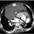
FIGURE 7.4 Pleural metastasis in a 16-year-old boy with testicular cancer. A: Contrast-enhanced axial CT image shows multiple enhancing pleural-based masses (arrows) with adjacent pleural effusion (E) and consolidation of the left lower lobe (asterisk). B: Contrast-enhanced axial CT image of the lower pelvis demonstrates a heterogeneously enhancing left testicular mass (arrow).


NORMAL ANATOMY

Pleura is a continuous surface epithelium that lines the thoracic cavity and the lungs. The visceral pleura adheres to the lungs and is in continuity with the parietal pleura, which covers the inner thoracic wall, diaphragm, and medial mediastinum. The visceral pleura invaginates into the interlobar fissures of the lung, as well as into accessory fissures if present (Fig. 7.6). In healthy children, the pleural membranes are typically 0.2 to 0.4 mm thick with ˜4 to 18 mL of physiologic fluid in the space, forming a layer 5 to 10 µm thick.3 Parietal and visceral pleura contain blood vessels and lymphatics. Visceral pleura is supplied by bronchial arteries, whereas parietal pleura is supplied by systemic arteries, including the phrenic artery and intercostal arteries. Venous drainage for the visceral pleura is to the pulmonary veins, whereas the venous drainage for the parietal pleura is to the intercostal and phrenic veins.


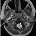
FIGURE 7.5 Pleural metastasis in a 15-year-old boy with synovial cell sarcoma. Axial T2-weighted MR image shows multiple low-signal pleural-based masses (asterisk) in the right hemithorax. (L, lung; E, pleural effusion.)

The pleural space is a potential space that normally contains a small amount of physiologic pleural fluid. Only the lymphatics of the parietal pleura communicate with the pleural space via 8- to 10-µm holes, which exude fluid into the pleural space (Fig. 7.7). This fluid is typically taken up by mediastinal lymph nodes, which drains into the thoracic duct and ultimately into the systemic venous system. Normal physiologic fluid is evenly distributed in the pleural space, decreasing friction between the visceral and parietal pleura during movement of the lungs against the chest wall with normal respiration.4 Pleura and the pleural space only become visible on imaging studies when an abnormality is present.


SPECTRUM OF PLEURAL DISORDERS


Pneumothorax

Pneumothorax is defined as air within the potential pleural space. Although there are various underlying etiologies of pneumothorax, they can be broadly categorized into either spontaneous (i.e., primary) or iatrogenic (i.e., secondary) pneumothorax. Spontaneous pneumothorax can occur in a previously healthy child resulting from spontaneous rupture of a preexisting bleb, usually located in the apex of the lung (Fig. 7.8), or may be a consequence of an underlying pulmonary disorder, such as asthma or cystic fibrosis. Secondary or iatrogenic pneumothorax can occur in the setting of a
recent intervention, such as a lung biopsy, thoracentesis, or mechanical ventilation.5 Iatrogenic pneumothorax can be also secondary to blunt or penetrating trauma, with or without an associated rib fracture (Fig. 7.9). Air can be introduced into the pleural space not only from an external wound but also secondary to pulmonary, esophageal, or tracheobronchial injuries.


FIGURE 7.6 Normal pleural anatomy. Schematic diagram of the lungs shows the parietal pleura outlining the chest wall and the visceral pleura overlying the lung as well as invaginating into the different fissures. The parietal and visceral pleural are continuous (pleural reflections) creating the potential pleural space.


FIGURE 7.7 Normal pleural anatomy. The lymphatics of the parietal pleura produce the majority of the pleural fluid, which is exuded into the pleural space via stomata. The mesothelial cells secrete a surfactant-like substance that lubricates the pleural cavity.


FIGURE 7.8 Spontaneous pneumothorax in a 15-year-old boy who presented with acute shortness of breath and right-sided chest pain. Coronal reformatted lung window CT image shows a large right pneumothorax with collapse of the right lung. Also noted are small blebs (arrow) at the right lung apex most likely the cause of this patient’s spontaneous pneumothorax.


FIGURE 7.9 Traumatic pneumothorax in an 8-year-old boy status post motor vehicle accident. Axial lung window CT image shows a left pneumothorax (asterisk). Opacities in the left lung are consistent with lung contusions with pneumatoceles (arrows) containing fluid (hemorrhage).

The incidence of spontaneous pneumothorax is ˜1.2 to 6 cases per 100,000 girls and 7.4 to 18 cases per 100,000 boys, with a mean age at presentation being 14 to 15.9 years in the pediatric population.5 The common demographic of pediatric patients with a spontaneous primary pneumothorax is tall, thin boys with low body mass index.6 Affected pediatric patients typically present with sudden-onset shortness of breath and unilateral chest pain. In the setting of thoracic trauma, one-third of thoracic injuries are associated with pneumothorax. A concerning adverse outcome of a traumatic pneumothorax is a tension pneumothorax, which occurs when the pleural defect acts as a one-way valve, allowing air to flow into the pleural cavity without escape. The increasing volume and accumulation of air in the pleural space cause mass effect on the adjacent structures, resulting in mediastinal shift to the contralateral side and possible vascular compromise of the heart and the great vessels (Fig. 7.10).5 Physical examination findings specific to tension pneumothorax include engorged neck veins and deviation of the airway to the contralateral side.

Standing AP or PA radiographs are an initial imaging method for evaluating pediatric patients clinically suspected of having pneumothorax. On frontal chest radiographs, pneumothorax typically presents as lucency and collapsed lung with visible pleural surface (Fig. 7.11). When the pneumothorax is localized in the subpulmonic region, lucency can be localized and projects over the lower lung zone, just above the diaphragm (Fig. 7.12). In the setting of both air and fluid in the pleural space, which is known as hydropneumothorax, the interface between the air and fluid can be seen on an erect radiograph as a horizontal line extending across the entire length of the hemithorax (Fig. 7.13). In critically ill pediatric patients who cannot tolerate standing, chest radiographs can be obtained in the supine position, which unfortunately decreases sensitivity for evaluation of pneumothorax. However, given air rises to the nondependent position, pneumothorax can be seen on a supine radiograph as global lucency in the chest without clear visualization of pleural line, especially in neonates and infants (Fig. 7.14). Lateral radiographs are not necessary as the initial evaluation; however, lateral decubitus radiographs can be useful in challenging cases particularly when the amount of intrathoracic free air is small. In severely ill
patients, a cross-table lateral radiograph or decubitus radiograph can be performed.

Only gold members can continue reading. Log In or Register to continue

Stay updated, free articles. Join our Telegram channel

Oct 13, 2018 | Posted by in PEDIATRIC IMAGING | Comments Off on Pleura

Full access? Get Clinical Tree

Get Clinical Tree app for offline access