Pleural Effusions

62
Pleural Effusions


Exudative versus Transudative Effusion on Computed Tomography


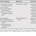
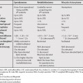
The most common cause of transudative effusion is congestive heart failure. The most common cause of exudative effusion is infection or pneumonia. In older patients, exudative effusions secondary to malignancy should be considered (Table 62.1). Thick enhancing pleural rind suggests exudative effusion. Pleural-based soft tissue nodules may be seen in malignant effusions on computed tomography (CT). Lung cancer and breast lymphomas are the cause of 75% of malignant effusions. A hematocrit effect (layering of blood products) should be looked for on the narrow mediastinal window to assess for hemothorax.


Mechanisms by which Malignancy Leads to Pleural Effusion


  • Direct

    • Pleural metastasis
    • Obstruction of pleural lymphatic vessels
    • Mediastinal lymph node involvement with decreased pleural lymphatic drainage
    • Chylothorax (thoracic duct obstruction)
    • Bronchial obstruction (decreased pleural pressures)
    • Pericardial involvement

  • Indirect

    • Hypoproteinemia
    • Postobstructive pneumonitis
    • Pulmonary embolism
    • Postradiation therapy


Only gold members can continue reading. Log In or Register to continue

Stay updated, free articles. Join our Telegram channel

Jan 10, 2016 | Posted by in MAGNETIC RESONANCE IMAGING | Comments Off on Pleural Effusions

Full access? Get Clinical Tree

Get Clinical Tree app for offline access
Table 62.1 The Appearance of Transudative versus Exudative Effusion on Computed Tomography