Fig. 25.1
Poliomyelitis. (a) Cervical plain MR imaging with sagittal SE sequence T2WI demonstrates continuous strip of high signal at the anterior spinal cord. (b) Transverse SE sequence T2WI demonstrates abnormal high signal at the ventral gray matter of anterior horn of C4–5 spinal cords (Reprint with permission from Malzberg et al. 1993)
Case Study 2
A male patient aged 28 years complained of low-grade fever as well as progressive weakness of lower limbs and left upper limb for 4 days. By cerebrospinal fluid examination, lymphocytes count increased, and levels of glucose and protein are normal. The patient reported no history of polio vaccination. His daughter had a recent history of peroral polio vaccination.
For case detail and figures, please refer to Haq A, et al. Arch Neurol, 2006, 63(5):778.
25.7.2 Skeletal System
25.7.2.1 Foot Deformity
Poliomyelitis can induce a variety of foot deformities, such as talipes equinovarus and talipes equinovalgus, talipes calcaneus, platypodia, and calcaneovarus deformity. The deformity can be unilateral or bilateral and singular or multiple.
X-Ray
It can be applied for examining the foot deformities as well as postoperational assessment.
1.
Talipes equinovarus
The astragalus is wide and flat, which is by anteroposterior X-ray demonstrated with the extension line of its central axis deviating far from the first metatarsal bone, while normally the line should penetrate through the first metatarsal bone. The calcaneus is short and wide, which shows introversion or upward shift. The navicular bone is wedge shaped. The introversion of propodeum into horseshoe-like appearance can be found, with depressed foot arch, metatarsal bones drawn together, dropping of the first metatarsal bone, semi-dislocation of the metatarsophalangeal joint, and obviously thickened soft tissue density shadow under the joint.
2.
Pes arcuatus
The instep is depressed like an arch, with shrinkage of both internal and external arches. The angle formed by lines connecting the lowest point of astragalus, the lowest contact point of calcaneus to the horizontal line, and the lowest contact point of the first metatarsal heat to the horizontal line is normally 113–130°, which is known as the internal arch. The angle formed by lines connecting the lowest point of calcaneal surface at the calcaneocuboid joint, the lowest contact point of the fifth metatarsal heat to the horizontal line, and the lowest contact point of calcaneus to the horizontal line is normally 130–150°, which is known as the external arch. The scaphoid bone is in wedge shape with its downward tip. The calcaneus and astragalus are morphologically normal, with normal locations. The metatarsal bones lean plantarly to form semi-dislocation of the metatarsophalangeal joint, with raised phalanx.
3.
Platypodia
The external arch of the foot is flattened, and the internal arch is pressed downward. The talus bone head, scaphoid head, cubitale, and cuboid bone shift downward, with the talonavicular joint developing into proliferative osteoarthropathy. The calcaneus is flattened with the valgus. Lateral X-ray demonstrates enlarged Boehler’s angle, which is an angulation by the longitudinal axis of talus and the longitudinal axis of calcaneus and is normally 25–50°.
4.
Calcaneovarus deformity
The stress lines of lower limbs of the patients shift forward while standing or walking. Lateral X-ray of the ankle joint demonstrates the presence of heel bone. However, the stress lines of the lower limbs normally pass through the talus center. By lateral X-ray of the ankle joint, the extension line of the tibial longitudinal axis passes through the talus ankle joint center. By measurement, both tibiocalcaneal angle and Bohler’s angle are enlarged, while normally the tibiocalcaneal angle, posterior angulation by the extension line of the tibial longitudinal axis and the calcaneus axis, is about 120°. Calcaneovarus deformity is commonly complicated by high arch deformity. Lateral X-ray of the foot demonstrates shrinkage of the internal arch and enlarged posterior arch (normally 13–16°). The characteristic anterior bone defect at the inferior tibia and shallowed ankle mortise can be observed, with elevated arch at the trochlea of astragalus and deepened depression at the dorsal talus neck.
5.
Talipes calcaneus
The heel is vertical, with obviously smaller angle between the heel and axis. The soft tissue at the heel is subject to obvious thickening, with raised toes.
Application of CT Scanning
By applying processing techniques after CT scanning, the foot deformities can be more favorably demonstrated.
25.7.2.2 Malformation of the Hip and Knee Joints
X-Ray
Sequelae of poliomyelitis possibly induce deformity of the hip and knee joints. In children, the transverse and longitudinal diameters of the femoral head, the distal femur of the knee joint, and the epiphyseal nucleus at the proximal tibia as well as the transverse diameter of metaphysic are smaller than normal. The development of the epiphysis is affected. The affected limb is shortened. The both ends of the bone are demonstrated with multiple growth disturbance lines. The deformity of hip joint is demonstrated as pelvic inclination, shallow acetabulum, dislocation of femoral head, and other deformities of femoral head, such as abduction, adduction, and rotation. The iliac wing, pubis, and ischium develop microgenesis and deformity, with narrowed or absent obturator foramen. The deformity of knee joint is demonstrated as knee flexion (with anterior flexion of the lower 1/3 of the femur and upper 1/3 of the tibia, hypertrophy of the femoral condyle, elevated anterior tibial plateau, and downward shift of the patella), genu recurvatum (with low and flat anterior tibial plateau, flat anterior femoral condyle, and no downward shift of the patella), and genu valgum (the outward open angle being smaller than 170° between the long axis of the tibia and the long axis of the femur, which is normally 170–179°).
CT Scanning
It can also be applied to demonstrate skeleton changes during the sequela stage of poliomyelitis. Post-processing techniques after CT scanning can more favorably demonstrate the complex deformities.
25.7.2.3 Osteoporosis and Muscular Atrophy
X-Ray and CT Scanning
During the sequela stage of poliomyelitis, in addition to limb deformities, osteoporosis of the affected limb can also be demonstrated with decreased and thin bone trabecula, thin bone cortex, decreased bone density, obvious growth disturbance line at the ends of long bones, and obvious white lines of epiphyseal trace at the metaphysis (Figs. 25.2 and 25.3).
MR Imaging
In the cases with muscular atrophy of the affected limb, MR imaging can also well demonstrate the amount and range of involved skeletal muscle, the decreased muscular volume of the affected limb, and the adipose substitution, which are nonspecific due to the commonalities with pathological skeletal muscles caused by other disease, such as cerebral paralysis and spinal muscular atrophy. But the lesions of poliomyelitis are typically distributed asymmetrically. MRS for patients with sequela of poliomyelitis demonstrates the presence or absence of intracellular adipose at the skeletal muscles which is related to the severity of paralysis. In the patients with serious paralysis, the intracellular adipose is demonstrated to be absent, with decreased or absent choline and creatine peak (Figs. 25.4 and 25.5). CT scanning can also demonstrate adipose substitution at the involved skeletal muscles of the affected limb. But the demonstrations are less favorable than those by MR imaging. By X-ray, the only demonstration is thinner soft tissue density shadow caused by muscular atrophy of the affected limb.
Case Study 3
A male patient aged 19 years developed poliomyelitis in childhood.
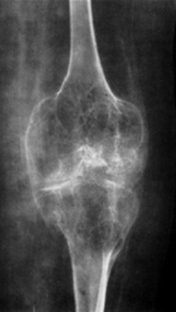
Fig. 25.2
Poliomyelitis complicated by bone lesions. X-ray demonstrates osteoporosis around the knee joint, narrow joint space, and hypertrophy of epiphysis as well as fine diaphysis of the femur and the fibula (Reprint with permission from Richardson et al. 1984)
Case Study 4
A female patient aged 34 years developed poliomyelitis at the age of 6 months, with paralysis of the right lower limb.
For case detail and figures, please refer to Liu T, et al. Chinese Modern Medication, 2008, 2 (6): 69. (In Chinese.)
Related posts:

Stay updated, free articles. Join our Telegram channel

Full access? Get Clinical Tree


