5 Porta Hepatis
LEARNING GOAL
 Identify and evaluate the vascular structures about the porta hepatis.
The porta hepatis can be a challenging region for the ultrasound novice. But by working systematically to learn this region, you should be able to identify and evaluate its vascular structures without difficulty.
Three vascular structures enter or leave the liver at the porta hepatis: the portal vein, the hepatic artery, and the common hepatic duct. The inferior vena cava runs distal to this triad (Fig. 5.1). The bile duct lies approximately on the longitudinal body axis. The hepatic artery and portal vein run almost parallel for a short distance, at a slight angle to the longitudinal body axis. The initial portion of the hepatic artery is almost perpendicular to this body axis (Fig. 5.2).
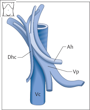
Fig. 5.1 The vessels of the porta hepatis: bile duct (Dhc), hepatic artery (Ah), portal vein (Vp), and vena cava (Vc). This diagram is familiar to you from anatomy. Take note of the following relationships: the bile duct and hepatic artery are anterior to the portal vein; the bile duct is lateral to the hepatic artery and portal vein.
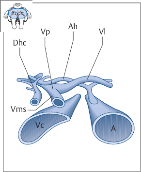
Fig. 5.2 Transverse section through the porta hepatis. Dhc = common hepatic duct, Vp = portal vein, Vms = superior mesenteric vein, Vl = splenic vein, Ah = hepatic artery, Vc = vena cava, A = aorta.
 Organ Boundaries: Identifying the Vessels in the Porta Hepatis
TIP
When defining the portal vein in the upper abdominal transverse scan, it is helpful to have the subject expand the abdomen.
The key sonographic landmark for the portal vessels is the portal vein itself. An upper abdominal transverse scan can define this vessel in its course anterior to the vena cava. It is helpful to have the subject take a deep breath, expanding the abdomen. Figure 5.3 shows the typical ultrasound appearance.
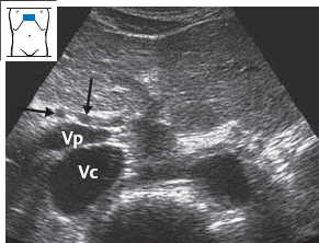
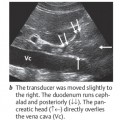
Fig. 5.3 Transverse scan at the level of the porta hepatis. You should positively identify two vessels at this level: the vena cava (Vc) posteriorly and the adjacent portal vein (Vp) anteriorly. The vascular sections in front of the portal vein are initially difficult to identify: the hepatic artery (↓) and common hepatic duct (→).
Vena cava and portal vein
These vessels can be identified by applying a simple technical principle. First confirm the identity of the vena cava.
Identifying the vena cava
Define the vena cava and portal vein in cross section as shown in Fig. 5.3. While watching the screen, rotate the transducer to a longitudinal scan over the vena cava so that you can positively identify that vessel. Then return to the initial position over the portal vein and vena cava.
Identifying the portal vein
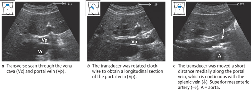
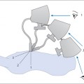
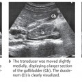
Confirm your identification of the portal vein by rotating the transducer clockwise from the transverse plane (Fig. 5.4a) until you see a longitudinal section of the portal vein (Fig. 5.4b). Move the transducer a short distance medially, and you will have no trouble recognizing the terminal portion of the splenic vein (Fig. 5.4c). Trace that vessel back to the portal vein.
Hepatic artery and bile duct
The main problem in examining the porta hepatis is in distinguishing between the hepatic artery and the bile duct. This should not be difficult, however, if you are familiar with the anatomy of the region.
Since the cystic duct cannot be positively identified with ultrasound, we will refer to the common hepatic duct and common bile duct collectively as the bile duct.
Recall that the hepatic artery curves across the vena cava to the celiac trunk, while the bile duct runs almost longitudinally, parallel to the vena cava, toward the head of the pancreas. This means that you can obtain a longitudinal section of the hepatic artery by rotating the transducer counterclockwise.
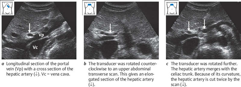
Identifying the hepatic artery
Place the transducer obliquely along the right costal arch, and image the portal vein in longitudinal section as described above under “Identifying the portal vein” (Fig. 5.5a). Now rotate the transducer counterclockwise to an upper abdominal transverse scan. You will see the hepatic artery just above the portal vein, coursing left toward the aorta (Fig. 5.5b). Trace the longitudinal section of the hepatic artery to the celiac trunk and then back to the porta hepatis (Fig. 5.5c).
Identifying the bile duct
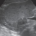
Image the portal vein again (Fig. 5.6a
Related posts:
Stay updated, free articles. Join our Telegram channel

Full access? Get Clinical Tree


