13 The Systematic Ultrasound Examination
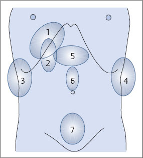
Now that you have learned the sonographic anatomy and interrelationships of the relevant organs, here are some guidelines on how to conduct a systematic ultrasound examination of the abdomen. Of course, the sequence in which the parts of the examination are performed will vary from one examiner to the next. The main thing for the beginner is to adopt a systematic routine that can be carried out within a certain period of time. In this system the abdomen is divided into seven topographic units:
1 Liver
2 Gallbladder and porta hepatis
3 Right kidney
4 Left kidney and spleen
5 Epigastrium and pancreas
6 Midabdomen
7 Lower abdomen
These units are shown schematically in Fig. 13.1 and are covered individually below.
Topographic Units
Shape
– Angle of inferior border
– Surface
Size
Structure
– Hyperechoic
– Pattern: coarse/fine
– Distribution
– Masses?
Vessels
– Hepatic veins
Diameter
Course
– Portal vein
Diameter
Course
Biliary tract
GALLBLADDER
Location
Size
Shape
Contents
Wall
Tenderness
Contracted?
Liver
Examination of the liver proceeds in three steps:
Survey in longitudinal scans.
Survey in transverse/oblique scans.
Survey in intercostal scans.
Reporting guidelines
The liver is:
 normal in size and shape;
 enlarged, diameter… cm on the mid clavicular line (MCL);
 reduced in size.
The echo pattern:
 is normal,
 is homogeneous,
 shows slightly/moderately/markedly increased echogenicity.
The angle of the inferior border is:
 sharp,
 slightly rounded,
 blunted.
The hepatic veins are:
 normal in appearance,
 rarefied,
 distorted.
No masses are seen.
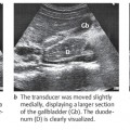
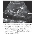
Gallbladder and porta hepatis
Examination of the gallbladder proceeds in three steps:
1 Survey in longitudinal scans.
2 Survey in transverse/oblique scans.
3 Lateral intercostal scans.
Examination of the porta hepatis proceeds in two steps:
1 Upper abdominal oblique scan (“portal scan”).
2 Subcostal oblique scan.
Reporting guidelines
The gallbladder:
 appears normal in size and shape,
 shows postprandial contraction.
Location
Shape
Size
Mobility
Parenchymal pelvic ratio
Parenchymal pattern
Renal pelvis
– Contents
– Origin of ureter
Demonstrable adrenal gland?
SPLEEN
Size
Shape
Hilum
Accessory
Spleen
Varices
A lumen is not seen.
The gallbladder is free of stones.
Several stones with acoustic shadows are noted: size … cm.
The gallbladder is folded over at the fundus (“Phrygian cap”).
The gallbladder wall is thickened.
The gallbladder is tender to local pressure.
Related posts:
Stay updated, free articles. Join our Telegram channel

Full access? Get Clinical Tree


