Chapter 40 Positron emission tomography (PET) scanning
Chapter contents
40.1 Aim
The aim of this chapter is to introduce the reader to the principle of positron emission tomography (PET) scanning as practised in diagnostic imaging. The assumption is made that the reader is familiar with the mechanisms of positron production from a radionuclide as described in Chapter 19 (Section 19.5.2.).
40.2 Revision of positron physics
As was discussed in Chapter 19, if a nucleus has too few neutrons for stability, it is possible for the nucleus to achieve a more stable configuration by the emission of a positron. Positrons are the antiparticles of electrons and the positron and the electron will interact (within a very short distance in tissue), annihilating each other and producing two photons of annihilation radiation. These photons each have an energy of 0.51 MeV (511 keV) and detection of these photons forms the basis of positron emission tomography. Each photon is produced at an angle close to 90° to the direction of travel of the positron (see Fig. 19.7).
40.3 Detection of positrons
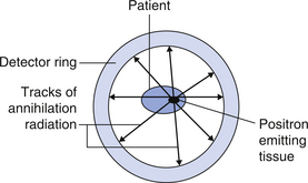
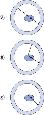
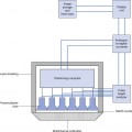
If a positron-emitting radionuclide is introduced into the patient, it can be labelled in such a way that it concentrates in specific structures. The number of positrons emitted by these structures can be related to the activity of the cells within the structure. If we surround the patient with a ring of positron detectors (see Fig. 40.1) then each annihilation radiation from the positron–electron interaction can be registered and the intersections of these points can be ‘back-projected’ to indicate the positron-emitting tissue in the patient.
