Presentation and Presenting Images
A 59-year-old female with a significant family history of breast cancer and a prior benign right excisional biopsy presents for routine screening mammography.
23.2 Key Images
23.2.1 Breast Tissue Density
There are scattered areas of fibroglandular density.
23.2.2 Imaging Findings
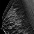
The imaging of the left breast is normal (not shown). The digital mammograms of the right breast shows a possible architectural distortion (circle) in the middle depth at the 11 o’clock location, 4 cm from the nipple ( ▶ Fig. 23.3 and ▶ Fig. 23.4). Scar markers (arrows) denote the scarring from the remote excisional biopsy. The tomosynthesis movie demonstrates that the possible architectural distortion is a summation artifact and the result of overlapping breast parenchyma.
23.2.3 BI-RADS Classification and Action
Category 1: Negative
23.3 Differential Diagnosis
Overlapping parenchyma: Although findings on the conventional mammogram suggested an architectural distortion, tomosynthesis demonstrated that the finding was created by overlapping parenchyma.
Radial scar: Tomosynthesis did not reveal a persistent architectural distortion nor mass.
Cancer: Cancers, particularly invasive lobular carcinomas, may present on one view only or as subtle findings. Tomosynthesis makes missing such a finding less likely than on conventional mammography alone.
23.3.1 Essential Facts
Lesion detection challenges due to overlapping breast parenchyma are a limitation to conventional two-dimensional (2D) analog and digital mammography.
In this case, if only 2D digital screening mammography had been performed, the patient would have been scheduled for diagnostic mammography and additional views, ultrasound, and possibly a biopsy. Obtaining the 2D digital mammography along with digital breast tomosynthesis (DBT) allowed for direct comparison between the 2D mammogram and DBT images.
The added DBT images demonstrate that the possible architectural distortion is a summation artifact. No further work-up or biopsy are needed. The patient can return to annual screening mammography, and the radiologist’s recall rate is reduced.
Overlapping breast parenchyma on mammography is one factor that limits interpretation, particularly in patients with denser breast tissue. Overlapping breast parenchyma may obscure cancers, resulting in missed cancer diagnoses. Conversely, overlapping parenchyma, or superimposed normal structures, may create pseudomasses, or summation artifacts, resulting in false-positive diagnoses and unnecessary biopsies.
23.4 Management and Digital Breast Tomosynthesis Principles
Breast tomosynthesis captures images of the breast at multiple angles during a short scan. The images are then reconstructed into thin slices (1-mm thick) that can be viewed individually or in a tomosynthesis movie. This essentially removes overlapping structures and/or superimposition of structures.
Many screening callbacks are performed to resolve findings secondary to overlapping parenchyma.
The addition of tomosynthesis to 2D digital mammography helps identify summation artifacts secondary to overlapping breast parenchyma, thereby decreasing the recall rate for diagnostic mammography and ultrasound and eliminating some unnecessary biopsies.
Tomosynthesis has been shown to reduce recall rates from 7 to 15%.
23.5 Further Reading
[1] Friedewald SM, Rafferty EA, Rose SL, et al. Breast cancer screening using tomosynthesis in combination with digital mammography. JAMA. 2014; 311(24): 2499‐2507 PubMed
[2] Rose SL, Tidwell AL, Bujnoch LJ, Kushwaha AC, Nordmann AS, Sexton RJr. Implementation of breast tomosynthesis in a routine screening practice: an observational study. AJR Am J Roentgenol. 2013; 200(6): 1401‐1408 PubMed
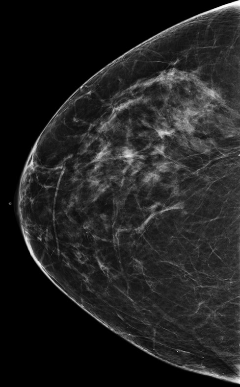
Fig. 23.1 Right craniocaudal (RCC) mammogram.
Related posts:


Stay updated, free articles. Join our Telegram channel

Full access? Get Clinical Tree


