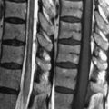
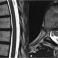
23 Posterior Fossa Congenital Malformations Chiari malformations are commonly encountered congenital abnormalities. Type 1 malformations are typically asymptomatic, although they may present with headaches, cerebellar signs, or lower CN symptoms. In addition to congenital etiologies, type 1 malformations may result from elevated intracranial pressure and conditions causing basilar invagination (e.g., Paget disease of bone). Figure 23.1A demonstrates the principle feature of a type 1 malformation—the herniation of wedge-shaped cerebellar tonsils (black arrow) below the foramen magnum. In this patient, the tonsils descend almost to the level of the posterior arch of C1. Herniation to the C1 level is seen in approximately two-thirds of patients; extension to the C3 level may occur in up to one-fourth of cases. Tonsils descending <5 mm below the foramen magnum are rarely of clinical significance. Figure 23.1B demonstrates another case of Chiari type 1 with the tonsils (black arrow) extending below the level of the posterior C1 arch. In this case, the malformation is associated with dilatation of the central canal of the cord, hydromyelia or (a more general term) syringohydromyelia (asterisk)—a longitudinally extending CSF-filled cavity commonly seen in this disorder. Hydromyelia demonstrates high SI on T2 and low SI on T1WI. Hydrocephalus is another frequent finding along with osseous abnormalities such as vertebral fusion, spina bifida occulta, and fusion of C1 to the occiput. The corpus callosum and quadrigeminal plate in Fig. 23.1A,B are normal, ruling out a Chiari type 2 malformation.
Related posts:


Stay updated, free articles. Join our Telegram channel

Full access? Get Clinical Tree


