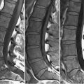
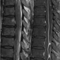
51 Primary Neoplasms As with spinal cord neoplasms in other regions, neoplasms of the lumbar spine are classified as extradural, intradural extramedullary, and medullary. If an extradural lesion contacts the cord, the subarachnoid space will be present at the interface. In the lumbar spine, extradural lesions predominantly consist of vertebral body tumors and metastatic disease (see Chapter 49). Neural sheath tumors and meningiomas comprise the major neoplasms in the intradural extramedullary space. The major nerve sheath tumors are schwannomas and neurofibromas, the imaging appearances of which were discussed in Chapter 39. These lesions are somewhat less common in the lumbar spine as compared with the thoracic. Frequently affecting the nerve roots within the inter-vertebral foramina in the cervical and thoracic spine, schwannomas within the lumbar spine may also involve the roots of the cauda equina. Such an appearance is demonstrated in the T2WI and CE T1WI of Figs. 51.1A,B, respectively. Schwannomas and neurofibromas are difficult to distinguish, even on MRI, although a heterogeneous appearance on T2WI (as in the rather lobulated lesion of Fig. 51.1) tends to favor the former. Schwannomas also occur peripheral to the nerve, tending to compress rather than enlarge it, more commonly exhibit cystic degeneration, and tend to be solitary rather than multiple. Figures 51.2A through 51.2C (T2WI, T1WI, contrast-enhanced T1WI) demonstrate a neurofibroma scalloping the posterior margin of the L2 vertebral body and widening the neural foramina in a patient with NF1. Based on the illustrations presented here, this lesion could represent a schwannoma or neurofibroma, however, in this case two additional enhancing intrathecal lesions were seen, favoring the latter. Although cystic degeneration is seen on T2WI (A) as an area of central high SI, a more characteristic appearance is that of a target-like configuration on T2WI with a bright rim surrounding a center of low SI. A nerve sheath tumor in this location may be distinguished from a foraminal disk herniation by the presence of bright contrast enhancement. A plexiform neuroma—pathognomonic for NF1—may occasionally be seen involving the sacral plexus as a bulky, multinodular, enhancing mass. Meningiomas are a further differential consideration with an intradural extramedullary mass. Unlike in the cervical spine, these tend to involve the posterolateral aspect of the canal. The widened subarachnoid space above and below the lesion in Figs. 51.3A,B,C localizes it to the intradural extramedullary space. As with the aforementioned lesions, this meningioma demonstrates isointensity to the cord on precontrast T2WI (A) and T1WI (B), most well-visualized on the former against the high SI CSF. (C
![]()
Stay updated, free articles. Join our Telegram channel

Full access? Get Clinical Tree


