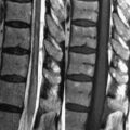
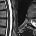
39 Primary Neoplasms Schwannomas and neurofibromas share a similar MRI appearance and are both common lesions in thoracic spine MRI. Schwannomas typically demonstrate heterogeneously high SI on T2WI with a moderate or low SI on T1WI. Foci of lower SI on T2WI may correlate with pathologically denser tissue in which there is a diminished amount of free water. In Fig. 39.1A, the extramedullary intradural mass—a location suggested by the relative broadening of the subarachnoid space at its margin with the tumor and the clear delineation of tumor from cord—compresses the spinal cord displacing it to the left. Both schwannomas and neurofibromas may occur less commonly extradurally or extend both intra- and extradurally leading to a dumbbell shape. As in the post-contrast T1WI of Figs. 39.1A,B these neoplasias often enhance heterogeneously, and small lesions may not be visible without contrast administration. The nonenhancing region of the tumor in Fig. 39.1B
![]()
Stay updated, free articles. Join our Telegram channel


Full access? Get Clinical Tree


