Chapter 34 Production of the digital radiographic image
Chapter contents
34.1 Aim
The aim of this chapter is to introduce the reader to the principles of digital imaging as practised in the radiological department. The assumption is made that the reader has read and understood Chapter 25 which dealt with the basic mechanisms of image production. The consequences of producing a digital image will be discussed in Chapter 36.
34.2 The digital image
The old method of producing a radiograph using film and intensifying screens is an example of an analogue image. The information (or data) it contains is represented by a range of continuously varying densities or shades of grey. If such an image is scanned as a series of horizontal lines and the densities plotted on a graph, we would see an appearance similar to that shown in Figure 34.1A (see page 252).
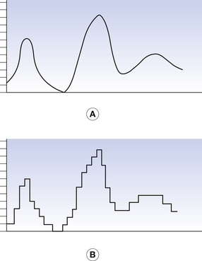
A digital image is divided into a series of small boxes called pixels, arranged in a series of rows and columns called a matrix (Fig. 34.2, see page 252). The density of each pixel has a numerical integer value. If we consider our initial radiograph, we could allocate the value 0 to the most dense value and 255 to the least dense value giving a digital scale of 256. Thus, any single pixel would have a discrete value between zero and 255 and our line would appear as a series of steps as shown in Figure. 34.1B. The smaller the image size and the larger the number of pixels in the image matrix, the better the spatial resolution of the image. If the pixels are too large, the individual pixels can be seen by the observer and distract from the image information.
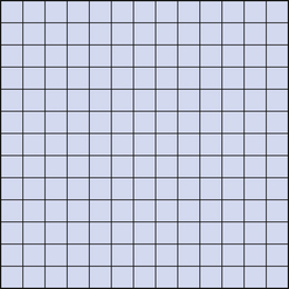
Figure 34.2 An example of the matrix used for a digitized image. Each box of this matrix is a pixel.