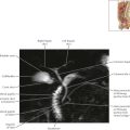
Prostate and Seminal Tract Axial 3

Normal anatomy
The seminal vesicles are paired accessory sex glands that produce and secrete fructose-rich seminal fluid, which is the major component of ejaculate volume. The detail of the convoluted, fluid-filled tubules that form the seminal vesicles is best depicted on T2-weighted MR images.

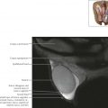
Prostate and Seminal Tract Axial 4

Diagnostic consideration
Denonvilliers’ fascia, also known as the rectoprostatic fascia, covers the seminal vesicles and the posterior aspect of the prostate gland, separating these structures from the rectum. Denonvilliers’ fascia helps limit the posterior spread of prostate cancer, making direct invasion of the rectum unusual.


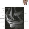
Prostate and Seminal Tract Axial 6

Normal Anatomy
The prostate gland is separated into a peripheral zone, central zone, and transitional zone, which constitute approximately 70%, 25%, and 5% of the prostate gland, respectively. The central and transitional zones are not well delineated on MRI and are collectively referred to as the central gland. Differentiation between the central gland and peripheral zone of the prostate is best appreciated on T2-weighted images, which reveal the posteriorly located, homogeneous, high signal intensity peripheral zone containing thin, linear, low signal intensity fibrous septa, in contrast to the anteriorly located, more heterogeneous, low intermediate signal intensity central gland.


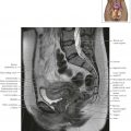
Prostate and Seminal Tract Axial 8