Scrotum and Testes


Scrotum and Testes Axial 1


Normal anatomy


On magnetic resonance imaging (MRI), the testicles (testes) are of uniform intermediate signal intensity on T1-weighted images, moderately high signal intensity on T2-weighted images, and enhance homogenously after intravenous contrast administration.


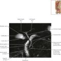
Scrotum and Testes Axial 2
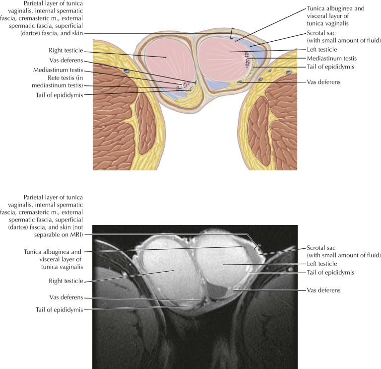
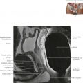
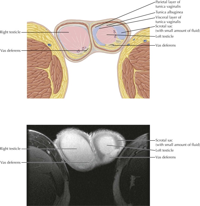


Normal anatomy


The testicles are composed of densely packed seminiferous tubules that converge posteriorly into larger ducts and drain into the rete testis at the testicular hilum. The mediastinum testis appears as a linear band of low signal intensity at the testicular hilum, where the ducts, nerves, and vessels enter and exit the testicle, while the rete testis has high signal intensity on T2-weighted MR images. It is important to be aware of the normal appearance and location of the mediastinum testis and rete testis so that these structures are not mistaken for pathology.
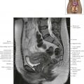


Scrotum and Testes Axial 3


Normal anatomy


The epididymides are isointense to slightly hypointense in signal intensity relative to the testicles on T1-weighted images, and hypointense to the testicles on T2-weighted MR images.


Scrotum and Testes Axial 4


Scrotum and Testes Axial 5


Normal anatomy


In men, the inguinal canal transfers the spermatic cord, which includes the vas deferens, testicular artery, pampiniform plexus of veins, and genital branch of the genitofemoral nerve, from the pelvic cavity to the scrotum.


Apr 4, 2019 | Posted by in RESPIRATORY IMAGING | Comments Off on Scrotum and Testes

Full access? Get Clinical Tree

Get Clinical Tree app for offline access