Pulmonary Capillary Hemangiomatosis
Jud W. Gurney, MD, FACR
Key Facts
Terminology
Rare cause of pulmonary hypertension due to proliferation of alveolar capillaries within lung
Imaging Findings
Enlarged pulmonary arteries + centrilobular ground-glass opacities
Top Differential Diagnoses
Pulmonary Venoocclusive Disease (PVOD)
Primary Pulmonary Hypertension (PPH)
Chronic Pulmonary Thromboemboli
Pathology
Some evidence that proliferation of thin-walled capillaries in PCH is histologic reaction to PVOD
Clinical Issues
Normal pulmonary capillary wedge pressure
Prognosis poor: Most patients die within 2 years of diagnosis
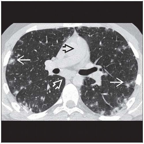 Axial NECT shows lobular nodular opacities with ground-glass halos  . Note the enlarged pulmonary artery . Note the enlarged pulmonary artery  and mildly enlarged lymph node and mildly enlarged lymph node  in this patient with pulmonary capillary hemangiomatosis. in this patient with pulmonary capillary hemangiomatosis. |
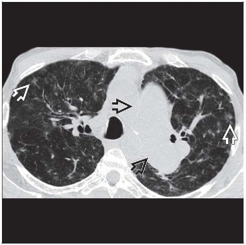 Axial NECT shows marked enlargement of the central pulmonary arteries
 and faint ground-glass opacities and faint ground-glass opacities  in pulmonary capillary hemangiomatosis. in pulmonary capillary hemangiomatosis.Related posts:Stay updated, free articles. Join our Telegram channel
Full access? Get Clinical Tree
 Get Clinical Tree app for offline access

|
