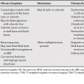
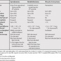
64 With the introduction of multidetector row computed tomography (MDCT), CT has been firmly established as the de facto first-line test for imaging patients with suspected pulmonary embolism (PE). Magnetic resonance imaging (MRI) for PE is indicated only in select cases, mostly with contraindication to iodinated contrast agents. Even in those cases, CT could be attempted using gadolinium DTPA (diethyl-enetriamine penta-acetic acid) as the contrast agent. CT, unlike conventional pulmonary angiography or a lung scan, can provide alternative diagnoses for patients’ signs and symptoms and reveal additional findings. The newer faster, multidetector CT with improved temporal resolution not only allows diagnosis of common noncardiac causes of acute pain such as acute or chronic PE (Table 64.1) or aortic dissection, but the same scan can be used to evaluate the heart and coronary arteries.
Pulmonary Embolism
Limitations of Computed Tomography
| CT Finding | Acute PE | Chronic PE |
|---|---|---|
| Filling defect | Central | Eccentric, mural |
| Lumen | Occluded | Nonoccluded |
| Embolus attenuation |


