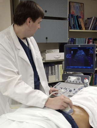
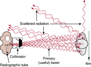
Radiation Safety and Protective Measures

CHAPTER 18


Radiation Safety and Protective Measures


The well known fact is that ionizing radiation, which is radiation that has sufficient energy to produce ions (e.g., x-rays), causes damage to living cells—damage that may be repaired, that may be permanent, or that may cause death to the cell. Therefore everyone involved in the medical application of ionizing radiation must have a basic knowledge of the many ways to minimize its lethal and sublethal effects.


Need for radiation protection


The two sources of ionizing radiation to which everyone is exposed are (1) natural environmental or background radiation and (2) human-made radiation. Examples of natural environmental or background radiation include cosmic radiation from the sun and stars; radioactive elements in the earth, such as uranium, radium, and thorium; and radioactive substances such as radiopotassium and radiocarbon, which are found in foods, drinking water, and the air. The amount of radiation that we receive from our natural environment depends to a great extent on where we live. One area of India has a high intensity of background radioactivity that gives the population 10 times more radiation than the average background radiation dose in the United States. People who live in high-altitude areas receive more cosmic radiation than those living in low-altitude areas; for example, the population in and around mile-high Denver receives more radiation than populations in or near sea-level coastal areas. Although background radiation varies from place to place, it accounts for more than one half of the exposure that the general public receives. Radiation has existed since time began. Diseases resulting from excessive radiation are not new either. The same kinds of harmful effects that radiation causes can also be caused by other agents, such as certain chemicals.


Human-made radiation sources include (1) fallout from nuclear weapons testing and effluents from nuclear power plants, (2) radioactive materials used in industry, and (3) medical and dental x-ray exposures. The use of medical and dental radiographs and radioactive materials to diagnose and treat disease accounts for 90% of the general public’s exposure to human-made radiation.


The possibility of radiation-induced injury was reported shortly after Roentgen’s discovery of x-rays in 1895. Since then, research, advanced technology, and the communications media have made society increasingly aware of the possible harmful effects of radiation; this awareness has lead to a belief that patient exposure to ionizing radiation must be kept to a minimum while obtaining optimal diagnostic information for the radiologist. Understanding the characteristics of x-radiation, its biologic effects, and the methods of reducing patient and operator exposure is the responsibility of the radiologic technologist.


Radiation Measurements


As awareness of the possible dangers of x-ray use increased, establishing a method of measuring its use became necessary. In the early days, persons who worked with x-rays used a unit of measure called the erythema dose. This unit was the amount of radiation required to turn the skin red, and its name was derived from the term erythema, which means redness of the skin. However, the erythema dose lacked preciseness and accuracy. A reliable instrument that measured the amount of ionization in gases was later developed, and the accuracy of this instrument allowed for the establishment of the unit of measurement known as the roentgen. This unit is the amount of ionizing radiation that produces, in 1 cubic centimeter of air, ions that carry 1 electrostatic unit of quantity of electricity of either positive or negative charge. The unit was named after the discoverer of x-rays, Wilhelm Conrad Roentgen. In 1938, the roentgen was adopted as the international standard measure of ionization in air. In 1956, another unit, called the radiation absorbed dose (rad), was established to measure the amount of radiation absorbed by a medium.


For introductory purposes, the long history of measurement standards and regulation, a brief overview of the National Council on Radiation Protection and Measurements, and the names and functions of other consumer protection agencies are presented.


Units of measurement are known as the roentgen (written as R in calculations), the rad, the roentgen-equivalent-man (rem), and the curie (Ci); the quantities associated with these units are exposure, absorbed dose, dose equivalent, and activity, respectively. The roentgen is a unit of exposure for x-rays and gamma rays. The rad is a unit of absorbed dose of any type of radiation. The rem is a unit that measures the biologic effect of x, alpha, beta, and gamma radiation on humans. The International System of Units (SI) uses coulomb*/kilogram (C/kg) in place of roentgen, gray (Gy) instead of rad, and sievert (Sv) rather than rem. For radiation protection from x and gamma radiation, 1 roentgen (C/kg) approximately equals 1 rad (Gy) or 1 rem (Sv). The Ci measures the amount of activity (known as radioactive disintegrations) that a radionuclide gives off. The unit of activity in the SI system is Becquerel (Bq); this measure is used in nuclear medicine studies with radionuclides, which are sometimes erroneously called radioactive isotopes (Table 18-1).


National Council on Radiation Protection and Measurements


In 1964 the Congress of the United States chartered the National Council on Radiation Protection and Measurements (NCRP) as a nonprofit corporation. The NCRP is composed of scientific committees the members of which are experts in their particular field or area of interest, and its primary function is to provide information and recommendations in the public interest about radiation measurements and protection. Another of its functions is to allow a pooling of resources from organizations to facilitate studies in radiation measurements and protection. A third function is to develop basic concepts about radiation protection and measurements and to develop the applications of these concepts. Last, the council makes a concerted effort to cooperate with international governmental and private organizations with regard to radiation measurements and protection.


The Radiation Control for Health and Safety Act of 1968 was an attempt to protect consumers from the hazards of radiation-producing electronic products. The U.S. Food and Drug Administration’s Bureau of Radiological Health is responsible for setting and regulating radiation performance standards that involve the manufacturing and assembly of radiation-producing electronic products, and it conducts ongoing research in an effort to minimize exposure to the patient, radiologic personnel, and the general public. The bureau also has a limited control program for radioactive materials that are not covered under the jurisdiction of the Atomic Energy Commission (AEC). Nuclear power production and the use of certain radioactive materials generally come under the control of the AEC. Environmental radiologic health protection is usually the responsibility of the Environmental Protection Agency.


Effective Absorbed Dose Equivalent Limits


The term effective dose equivalent (EDE; the absorbed dose multiplied by the appropriate quality factor and measured in rems) limit is in adherence to the radiation protection guides. The philosophy underlying the establishment of dose limits is twofold: The first premise is the no-threshold concept, and the second is the risks-versus-benefits relationship. Simply stated, the no-threshold concept is the belief that no known level exists below which adverse biologic effects may occur. This theory has created a great deal of controversy because it has not been proven conclusively; it is backed primarily by the observation of clinically induced irradiation to animals and extrapolation of high-dose irradiation received by atomic bomb survivors. If the assumption is that any amount of radiation can possibly cause deleterious effects to humans, then all radiation must be used prudently for the benefit of all concerned. The phrase, as low as reasonably achievable (ALARA), is the basis for the NCRP establishment of policies and procedures for radiation exposure.


The NCRP states: “The primary goal is to keep radiation exposure of the individual well below a level at which adverse effects are likely to be observed during his lifetime. Another objective is to minimize the incidence of genetic effects” (NCRP Report 34). Whenever physicians order a radiographic procedure, they must weigh the benefits to be obtained against the risk of the exposure.


Although every effort should be exerted to keep the dose of radiation at the lowest possible level for people who are well, this effort should not be a deterrent for the use of x-radiation for the detection and identification of disease processes in patients who are injured or ill, provided this use is performed by physicians and radiologic technologists who are trained and experienced in making such examinations.


Dose limits are categorized into two groups. Radiation workers, who are expected to receive radiation exposure during normal occupational activities, are limited by a maximal EDE of 5 rems per year. The general public is protected by a dose limit of 0.5 rem per year, which is one tenth of the total body limit for occupationally exposed individuals.


Students who are younger than 18 years and who are exposed to radiation during educational activities should receive no more than a 0.1-rem whole-body dose in 1 year. This EDE limit is based on the laws regarding minors. Thus this limit is the same for students younger than 18 years as it is for the general public. Minors employed in radiation areas are also included in these guidelines. After the age of 18, the student or employee is classified as an occupational worker (Table 18-2).


The total permissible dose to a pregnant woman should be no more than 0.5 rem because of the susceptibility of the developing embryo or fetus to the harmful effects of radiation. Authorities further suggest that the rate of exposure be controlled by specifying that the dose equivalent should not exceed 0.5 mSv (0.05 rem) in any given month. In fact, postponing any radiation exposure is advisable during the entire gestation period or to use another imaging modality, such as ultrasonography, to gather information for diagnosis (Fig. 18-1).


In summary, the radiologic technologist is now aware of the standard known as the EDE for occupational workers and that a dose limit has been established for the general population. Preventing the embryo or fetus from being exposed to any unnecessary radiation exposure is of primary concern.


The no-threshold concept and risks-versus-benefits relationship should always be kept in mind when considering the use of ionizing radiation in patient diagnoses. The information gained from a diagnostic radiograph should be far more beneficial than the possible risks incurred by exposure of the patient to ionizing radiation.


Interaction of x-rays with matter


An atom is the smallest part of an element and is made up of a nucleus surrounded by electrons. X-rays are packets of energy called photons, which have the ability to knock electrons out of their orbit; this action creates electrically charged ions. When x-rays pass through matter, this process of ionization results in a transfer of energy. The x-ray photons can be absorbed or scattered by the medium with which the photons interact or pass directly through the medium without any interaction taking place (Fig. 18-2).


The three main types of photon interactions that are important to radiology are (1) photoelectric effect, (2) Compton scatter, and (3) pair production.


Mar 2, 2016 | Posted by in GENERAL RADIOLOGY | Comments Off on Radiation Safety and Protective Measures

Full access? Get Clinical Tree

Get Clinical Tree app for offline access