Radioembolization for Hepatocellular Carcinoma


Radioembolization for Hepatocellular Carcinoma


Eleni Liapi and Jean-François H. Geschwind


Introduction


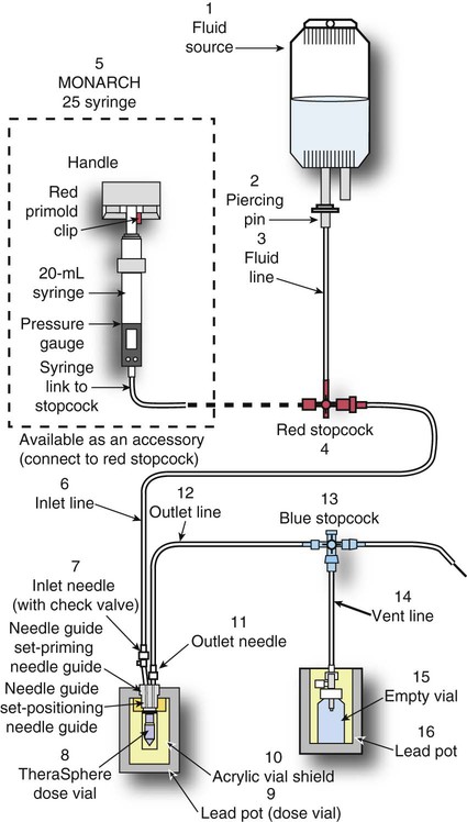
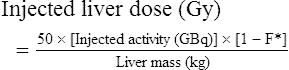
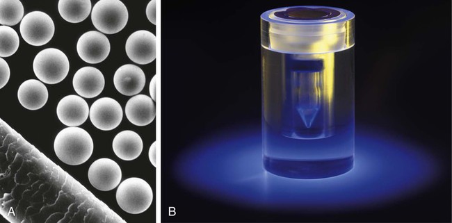
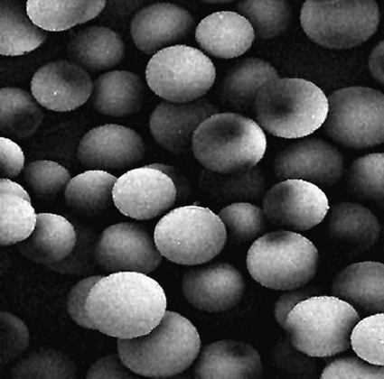
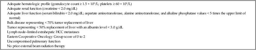
Conventional external radiotherapy has been limited and unsatisfactory for hepatocellular carcinoma (HCC), primarily because the liver has a low irradiation tolerance.1 The introduction of three-dimensional conformal radiotherapy (3D-CRT) and other computerized treatment planning techniques have significantly increased the use of radiation therapy in patients with unresectable HCC; however, radiation-induced liver disease (RILD) remains a major complication even when using 3D-CRT.2,3


Intraarterial injection of radioembolic microspheres into the tumor vessels has evolved as a promising answer to the challenge of delivering high-dose radiation to liver tumors while limiting dose to the uninvolved liver. By exploiting HCC’s preferential blood supply by the hepatic artery, radioembolization combines delivery of internal radiation to the tumor and concomitant microembolization of small intratumoral blood vessels. This chapter is focused on the two most commonly employed radioembolization methods for palliation of unresectable HCC: yttrium-90 (90Y) microsphere embolization and iodine-131 (131I)-Lipiodol embolization.


Yttrium-90 (90Y) Radioactive Microspheres


Clinical Relevance


The treatment of unresectable liver cancer with intraarterial injection of ceramic 90Y microspheres was first introduced in the 1960s.4,5 Early studies focused mainly on treating colorectal liver metastases, but further experience and encouraging results led to the application of 90Y microsphere embolization in patients with HCC.68 Currently, there are two types of commercially available radioactive microspheres:


They both contain 90Y as the active particle but differ in the type of carrier. 90Y can be produced by bombardment of stable 89Y with neutrons in a nuclear reactor. This radioactive product is a pure β emitter (937 kiloelectron volts [keV]) that decays to stable 90Zr, with a half-life of 64.2 hours. Emitted electrons have an average tissue penetration of 2.5 mm (effective max. 10 mm). One gigabecquerel (27 millicuries [mCi]) of 90Y per kilogram of tissue provides a dose of 50 Gy.


90Y glass microspheres (TheraSphere) are non-biodegradable insoluble microspheres that contain 90Y in a glass matrix. 90Y cannot leak out from this glass matrix. TheraSphere microspheres have a mean diameter of 25 ± 10 µm and 1 mg contains between 22,000 and 73,000 microspheres (Fig. 65-1). TheraSphere is supplied in 0.6 mL of sterile, pyrogen-free water contained in a 1-mL V-bottom vial secured within a clear acrylic vial shield. TheraSphere is available in six dose sizes: 3 GBq (81 mCi), 5 GBq (135 mCi), 7 GBq (189 mCi), 10 GBq (270 mCi), 15 GBq (405 mCi), and 20 GBq (540 mCi). Each dose of 90Y microspheres is supplied with an administration set that facilitates infusion of the microspheres from the dose vial (see later discussion). The intended dose of radiation to the targeted area ranges between 125 and 150 Gy (12,500-15,000 rads). Following intraarterial injection, TheraSphere microspheres embolize at the arteriole level. Histologic studies have shown increased accumulation of 90Y microspheres along the vascular periphery of the hepatic tumor, and up to 50 or 60 times more than in normal liver parenchyma.9 After lodging in the distal arteriolar circulation, the microspheres emit β radiation that penetrates tissue a maximum effective 10 mm, thereby sparing normal liver parenchyma beyond this limit. Radiation essentially ceases 10 days after embolization, but even before that it poses no threat to others.


Utilization of 90Y glass microspheres for intraarterial treatment of hepatic malignancies was initially approved in Canada in 1991. In 1999, a Humanitarian Device Exemption (HDE) was granted by the U.S. Food and Drug Administration (FDA) for treatment of unresectable HCC with 90Y microspheres, and their clinical use was initiated in the United States; some 800 patients with HCC have been treated since then.


The second type of 90Y microspheres (SIR-Spheres) is biocompatible, non degradable, and resin-based, with a diameter of 29 to 35 µm (Fig. 65-2). SIR-Spheres have an average activity of 40 Bq per sphere and can be suspended in sterile water and contrast media to the desired total activity. Compared with glass microspheres (specific activity of 2467 Bq per glass microsphere), resin microspheres have much lower specific activity per sphere. SIR-Spheres were granted premarket approval by the FDA in 2002 for treating unresectable metastatic liver tumors from primary colorectal cancer, in conjunction with adjuvant floxuridine-based chemotherapy administered via the hepatic artery. Most clinical trials for treatment of unresectable HCC with SIR-Spheres have been conducted in Australia, Hong Kong, and Europe.1012 Since the radioactive element is the same as that of glass microspheres, tissue penetration and decay characteristics are identical. Characteristics of the two devices are compared in Table 65-1. The radioactivity of 90Y delivered is dependent on the volume of liver and adjusted for shunting to the gastrointestinal (GI) tract and lungs based on estimates of flow from a technetium-99 (99Tc)-macroaggregated albumin scan.


Patient Selection and Preparation


In an institutional setting, a multidisciplinary panel consisting of interventional radiologists, medical and surgical oncologists, radiation oncologists, hepatologists, pathologists, and transplant surgeons may review patients’ eligibility for treatment with 90Y microspheres. Pretreatment evaluation always includes a routine clinical history, physical examination, complete blood cell count, blood biochemical analysis (including liver and renal function), and an α-fetoprotein assay. Selection criteria are similar to those for transcatheter arterial chemoembolization (TACE) and are listed in Table 65-2. Functional status is assessed by the Eastern Cooperative Oncology Group (ECOG) performance status. Okuda stage and Child-Pugh score should also be obtained before treatment. Pretreatment imaging may include a triple-phase contrast-enhanced spiral computed tomography (CT) scan of the abdomen, chest, and pelvis and/or contrast-enhanced magnetic resonance imaging (MRI) of the abdomen and pelvis to identify extrahepatic disease and calculate tumor and liver volumes. At our institution, the standard imaging workup includes a baseline gadolinium-enhanced MRI scan of the liver and abdomen, with diffusion/perfusion sequences for thoroughly assessing baseline tumor imaging characteristics as well as disease status.


Prophylactic therapy with gastric acid inhibitors on the day of treatment is highly recommended and has resulted in substantial reduction of associated GI symptoms.


Preoperative Planning


Pretreatment Visceral Angiography and 99mTc-Labeled Macroaggregated Albumin Injection


The purpose of performing baseline celiac and hepatic angiography is to define the vascular anatomy, plan a tailored treatment, and detect possible extrahepatic shunting. Prophylactic coil embolization of the gastroduodenal artery (GDA)—and/or any other collateral vessel or gastric variant (e.g., right gastric artery and its pancreaticoduodenal branches) that may result in microspheres being lodged into the GI area—is of no clinical consequence and highly recommended, since nontargeted delivery of 90Y microspheres to the GI tract can cause substantial morbidity. If the radioactive microspheres are planned to be injected from either the right or left hepatic artery (lobar treatments) and the tip of the catheter is placed far enough from the origin of the GDA, prophylactic GDA occlusion may not be necessary.


Accessory branches that contribute to the tumor vascular bed should also be readily identified because more treatments may be necessary, and the amount of radioactivity to be delivered must be calculated to include the relative contribution of each vessel to the liver volume to be treated. For instance, in the presence of a middle hepatic artery, which usually arises from the right hepatic artery, delivery of 90Y to the segment supplied by the middle hepatic artery may be precluded despite accurate catheter placement, given the flow dynamics. Therefore, three treatments (one each to the right, middle, and left hepatic arteries) may be necessary to cover the entire liver. Another example might include a patient with an accessory right hepatic artery, necessitating a third treatment (left hepatic [segments II-IV], right hepatic [segments V/VIII], and accessory right hepatic [segments VI/VII]).


Direct arteriovenous intratumoral shunting is common in HCC, so 90Y microspheres may shunt to the lungs, resulting in radiation-induced pneumonitis, with significant morbidity and possible mortality when the total lung dose approaches 30 to 50 Gy. Assessment of possible shunting to the lungs is therefore crucial before initiating treatment. A metastable technetium-99 (99mTc) macroaggregated albumin (99mTc-MAA) scan is performed after injecting 99mTc-MAA through the relevant hepatic artery (where treatment will be targeted) at the time of the pretreatment angiogram to calculate the percent radiation that might go to the lungs. The size of these albumin microspheres is 30 to 50 µm, which closely resembles the size of 90Y microspheres, so their injection may be similar to the distribution of 90Y microspheres. If activity is noted in the lungs, a shunt fraction is calculated as the ratio of the lung counts to the total counts. 99mTc-MAA scans cannot effectively demonstrate flow to the GI tract, and this drawback has been attributed to the much lower density of MAA particles (≈1.3 g/cm−3) than glass microspheres (3.7 g/cm−3). Overall, 99mTc-MAA provides a better simulation for resin-type microspheres (density 1.6 g/cm−3) than the glass type.


Radiation Dosimetry


It bears noting here that patients in whom the hepatopulmonary shunt fraction is greater than 10% of the injected dose with glass microspheres and greater than 20% with resin-based microspheres, or in whom the shunt fraction indicates potential exposure of the lung to an absorbed radiation dose of more than 30 Gy should not be considered for treatment with 90Y microspheres (Fig. 65-3).


As mentioned earlier, cross-sectional imaging (CT and/or MRI) is necessary to calculate liver volumes to assess the final amount of radioactivity to be delivered to the tumor site. For maximal effect on tumors, a dose of 100 to 150 Gy should be delivered to either lobe. Therefore, calculations of dose are based on the assumptions of a nominal target dose of 150 Gy/kg and a uniform distribution of microspheres throughout the liver volume. It is important to keep in mind that the dose is based on liver volume rather than tumor volume. The amount of radioactivity required to deliver the desired dose to the liver is calculated using the following formula:


Treatment activity required(GBq)=[Desired dose (Gy)]×[Liver mass (kg)]50


image


The liver mass is determined by measuring liver volume (mL) and using a conversion factor of 1.03 g/mL.


Calculation of the liver dose (Gy) delivered after injection is provided by the following formula:


Injected liver dose (Gy)=50×[Injected activity (GBq)]×[1F*]Liver mass (kg)


image


where *F is the fraction of injected activity localizing in the lungs, as measured by 99mTc-MAA SPECT (single-photon emission computed tomography).


Percentage of dose delivered to the patient can be calculated based on ion-chamber radiation detector measurements of the dose prior to administration, compared to measurements of the waste after administration. Before administration, the acrylic shield containing the dose is measured at a distance of 30 cm from the detector. After administration, the waste container inside the beta shield is measured at a distance of 30 cm from the detector at four rotational positions, and these four measurements are averaged. The percentage of dose delivered to the patient can be calculated using the following equation:


Percentage ofdose delivered (%)=[1Waste measurement after administrationDose vial measurement before administration]×100


image


Note that the calculation of the dose to be delivered with the 90Y resin microspheres is based on the extent of tumor involvement in the liver volume rather than on the assumption of a uniform distribution of radioactivity within the treated area.


Two methods are currently used to calculate the actual dose with resin microspheres: the body surface area (BSA) method and an empirical one. The empirical method uses the percentage of liver tumor involvement to calculate the recommended dose, and this dose can further be decreased, depending on the degree of arterioportal shunting on the 99mTc-MAA scan. According to the BSA method, the dose may be based on the BSA and the percentage of tumor involvement of the liver. The BSA is calculated in square meters as: BSA = 0.20247 × h0.725 × w0.425, where h is height in meters, and w is weight in kilograms. The percentage of tumor involvement of the liver (TI) is calculated as TI = (TV × 100)/(TV + LV), where TV is the volume of the tumor, and LV is the volume of the liver. The dose, then, may be calculated using the following equation:


Aresin=(BSA0.2)+(TI/100)


image

where Aresin is the activity of the 90Y content of the resin microspheres (in GBq).


Dec 23, 2015 | Posted by in INTERVENTIONAL RADIOLOGY | Comments Off on Radioembolization for Hepatocellular Carcinoma

Full access? Get Clinical Tree

Get Clinical Tree app for offline access