Fig. 1
Scheme. Anatomy of the different retroperitoneal spaces, the anterior pararenal space (brown), the perirenal space (yellow), the posterior pararenal space (violet), and the properitoneal compartment (dark violet)
The renal fasciae, namely, the anterior and posterior renal fasciae, represent the fundamental anatomical structures for the division of the retroperitoneal space, and they are clearly visible on computed tomography (Fig. 2) and in magnetic resonance imaging (Fig. 3). The renal fasciae are usually not thicker than 3 mm. If renal fasciae appear thicker than 3 mm, this is often due to a retroperitoneal space disease, corresponding mainly to acute pancreatitis (Fig. 4), renal pathologies, and abdominal aorta aneurysm rupture. The anterior pararenal space is limited anteriorly by the posterior sheet of the parietal peritoneum and posteriorly by the anterior renal fascia known also as Gerota’s fascia. Superiorly, it extends to the dome of the diaphragm and hence to the mediastinum and, inferiorly, communicates with the pelvis and the posterior pararenal space (Burkill and Healy 2000). It contains the pancreas, the second (descending), third (transverse), and fourth (ascending) loops of the duodenum, and the descending and ascending colon tracts. The ascending and descending colon are retroperitoneal and not abdominal organs because they do not have a meso formed by the peritoneal folds. The right and left sides of the anterior pararenal space communicate across the midline.
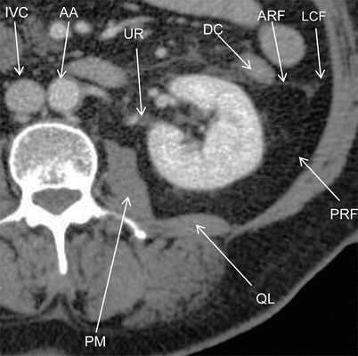
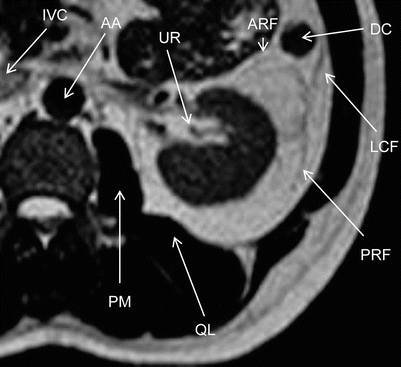
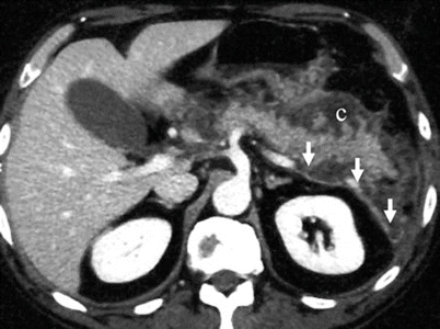
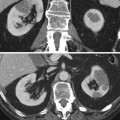
Fig. 2
Computed tomography. Anatomy of the retroperitoneal space. AA abdominal aorta, ARF anterior renal fascia, DC descending colon, IVC inferior vena cava, LCF lateroconal fascia, PRF posterior renal fascia, PM psoas muscle, QL quadratus lomborum muscle, UR ureter
Fig. 3
Magnetic resonance imaging, T2-weighted sequence. Anatomy of the retroperitoneal space. AA abdominal aorta, ARF anterior renal fascia, DC descending colon, IVC inferior vena cava, LCF lateroconal fascia, PRF posterior renal fascia, PM psoas muscle, QL quadratus lomborum muscle, UR ureter
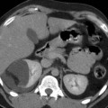
Fig. 4
Computed tomography. Transverse plane. Diffuse thickening of the anterior renal fascia (arrows) due to a collection located in the anterior pararenal space (c) due to a necrotic-hemorrhagic pancreatitis
The perirenal space (Figs. 5 and 6) is a retroperitoneal space that is limited anteriorly by the anterior renal fascia and posteriorly by the posterior renal fascia (Zuckerkandl’s fascia). Raptopoulos et al. (1986


Related posts:


Stay updated, free articles. Join our Telegram channel
Full access? Get Clinical Tree
