, Valery Kornienko2 and Igor Pronin2
(1)
N.N. Blockhin Russian Cancer Research Center, Moscow, Russia
(2)
N.N. Burdenko National Scientific and Practical Center for Neurosurgery, Moscow, Russia
In most cases, metastatic tumors classified as “rare forms of cancer” also did not show any specific diagnostic characteristics that would differentiate them from other metastases, but we find we have to present this small group of cases.
25.1 Cancer of the Parotid Salivary Gland
The most common tumor of the salivary glands is a pleomorphic adenoma—a benign mixed tumor consisting of both epithelial and mesenchymal tissues—and accounts for about 70% of all tumors of the salivary glands (Gnepp et al. 2001; Som et al. 2003). Most of these tumors are located laterally to the area of the facial nerve. Mostly, these are well-separated painless tumors; they occur in middle-aged patients. Malignant forms of ductal carcinoma are the most common malignant variant of salivary gland tumors. In 25% of cases, if untreated, they can metastasize with involvement of meninges (Thackray and Lucas 1983; Peel and Gnepp 1985; Som et al. 1988; Olsen and Lewis 2001). Malignant neoplasms are accompanied by severe pain and are identified in the elderly; infiltrative spread to the salivary gland tissue is possible. The 5-year survival is only about 50% (Som and Brandwein 2003), with the cause of death being metastases, including to the brain. The brain involvement is combined with involvement of the lymph nodes in the neck. Since the spread of the tumor occurs mainly by a hematogenous route, distant metastases are found in 44% of cases (Olsen and Lewis 2001). They usually affect the lungs, pleura, kidneys, and choroid. We observed three cases of intracranial metastases of the parotid gland tumor.
Metastases of the parotid gland cancer in the brain are often cystic, with the solid component rapidly accumulating the contrast agent. No reliable differential diagnostic characteristics have been identified using additional methods. No hemorrhages were observed (Figs. 25.1 and 25.2).
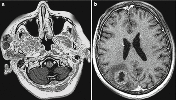
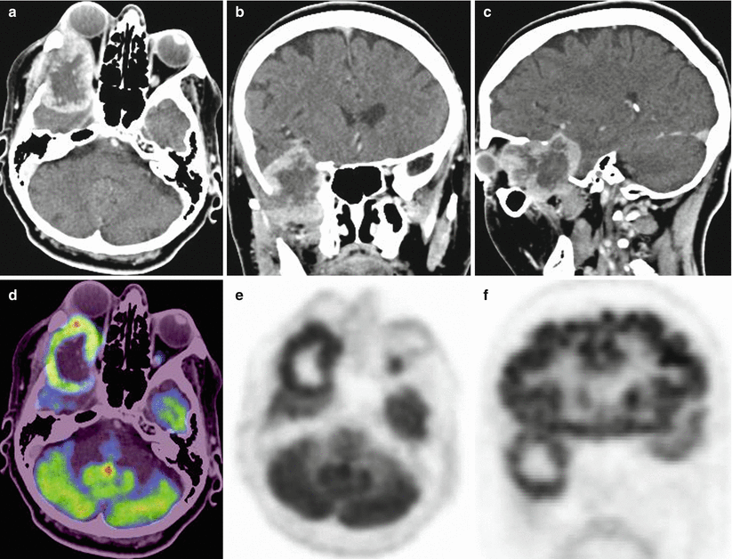

Fig. 25.1
Metastases of parotid gland cancer. A two-level study. On T1-weighted MRI with contrast enhancement, multiple tumor lesions are identified in the soft tissues of the right malar area (a). In the right parietal lobe region, there is a solitary rounded lesion that intensely accumulates the contrast agent of in the form of ring enhancement (b)

Fig. 25.2
A metastasis of parotid gland cancer in the brain. In the basal right temporal region, there is a lesion destroying the adjacent bone structures and extending to the region of the right orbit, infratemporal fossa, as well as intracranially, deforming the temporal lobe and shifting it upward (a–c). On a PET/CT study with 18F-FDG, there is an increase in SUV along the contour of the tumor (d–f)
25.2 Neuroendocrine Tumors (NETs)
Neuroendocrine tumors (NETs) heterogeneously group of epithelial tumors developing from APUD system cells, characterized by a positive immunohistochemical reaction to specific markers (chromogranin A, synaptophysin) and the ability to produce a variety of peptide hormones and biogenic amines. Standardized incidence rates for NETs in different countries vary within 0.71–1.36 per 100000 patient-year (Gulec et al. 2002). The most common site of NETs is the gastrointestinal tract (73.7%) and the bronchopulmonary system (25.1%). Within the gastrointestinal tract, most tumors are located in the small intestine (28.7%), the appendix (18.9%), and the rectum (12.6%). The overall 5-year survival of patients with NETs, regardless of their site, is 67.2–82% (Bektas et al. 2002).
NET metastases in the brain, including those of intracranial nature (pituitary tumors), are extremely rare (our clinical material includes two cases, one of which is presented below).
In most cases, NETs have no clinical symptoms to complications or to the development of carcinoid syndrome. Therefore, in most cases, a primary tumor and metastases are difficult to diagnose. The degree of differentiation of APUD cells and their functional activity and synthesis of hormones affect the rate and characteristics of tumor growth and its ability to metastasize. The principal method of treatment for NETs of the abdominal cavity and retroperitoneal space is surgery. Most authors have quite a unanimous opinion on the treatment strategy: a local excision can be performed for tumors smaller than 2.0 cm; radical operations are performed for larger tumors (subtotal resection of the stomach, small intestine resection, and hemicolectomy (Neumann et al. 2002; Soga 2003)). For conservative treatment, hormonal and chemotherapeutic drugs are used (6-methylprednisolone, ACTH, Endoxan, 5-fluorouracil). The effect of their use is low and short term.
Today, there are various methods for NET diagnosis: CT, MRI and ultrasound methods; lately, radioisotope methods are widely used—SPECT/CT with 111In-octreotide and PET/CT with 68Ga-DOTA (TOC, TATE, NOC). These drugs allow to evaluate the tumor receptor activity; therefore, they are highly specific in the diagnosis of NETs (Fig. 25.3).
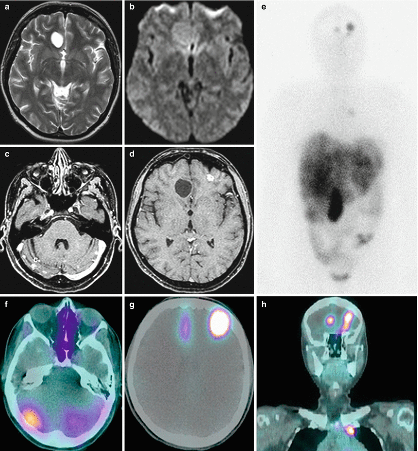

Fig. 25.3
Multiple neuroendocrine tumor metastases in the brain. Sub- and supratentorially, there are multiple lesions with various sizes. In the right frontal region, there is the largest cystic lesion with an increased signal on T2-weighted MRI (a), a moderately high signal on DWI MRI (b), that weakly accumulates the contrast agent along its contour (d). Small metastases in the left frontal region and in the hemispheres of the cerebellum have a mainly solid structure and intensely accumulate the contrast agent (c, d). There is no perifocal edema. A radionuclide study with 111In-octreotide on whole-body scans in the frontal projection (e) shows lesions with increased RP accumulation in the frontal areas of the brain, the hilum of the left lung, and the liver. On SPECT images in the axial (f, g) and frontal (h) projections, the cystic lesion in the right frontal region is characterized by a smaller area of increased RP accumulation than the small solid lesion in the left frontal region. Multiple pinpoint metastases in the cerebellum exhibit diffuse accumulation. A tumor site in the upper mediastinum is additionally identified (h) (the image is courtesy of Professor Shiryaev, S.V.)
25.3 Sarcoma (Fibrosarcoma)
Sarcoma (fibrosarcoma) is a malignant tumor of soft tissues that is formed from the immature fibrous connective tissue. It is often located in the muscles of extremities (hip, shoulder) or torso. Brain sarcomas are quite rare, occur with a frequency of up to 2%, and are usually located in the craniofacial area. There are infantile fibrosarcoma (in children under 10 years of age) and adult fibrosarcoma (common in children older than 10 years of age and in adults, more often at the age of 40–55 years old) (Toro et al. 2006).
Brain sarcoma has all the properties of a malignant tumor—aggressive rapid growth and the ability to grow into normal tissues and metastasize. Sarcomas are characterized by predominantly hematogenous metastasizing in 90% (Friedrichs et al. 2006), with not less than two third of patients having affected the lungs, less frequently bones, brain, pancreas, liver, and kidneys (Ottaiano et al. 2005). Warren and Meyer (1938) and then a number of other authors (Taylor and Nathanson 1942; Willis 1952; Pack and Ariel 1958) suggested the possibility of lymphatic metastasizing of sarcomas; however, it accounts for no more than 10% (Behranwala et al. 2004; Yanagawa et al. 2014). In our material, sarcomas are presented by two cases of intracranial metastases.
CT and MRI manifestations of craniofacial sarcoma are indistinguishable from those other malignancies growing in this area. By the time of primary diagnosis, the tumor is usually advanced.
Stay updated, free articles. Join our Telegram channel

Full access? Get Clinical Tree


