, Valery Kornienko2 and Igor Pronin2
(1)
N.N. Blockhin Russian Cancer Research Center, Moscow, Russia
(2)
N.N. Burdenko National Scientific and Practical Center for Neurosurgery, Moscow, Russia
Hemangioblastoma belong to rare, benign tumors, whose histological origin is still debated. It is believed that hemangioblastoma (HMB) occurs in 1–2.5% of all intracranial tumors. The proportion of childhood tumors accounts for about 20% of all Hemangioblastoma. The tumor is detected as one of the manifestations of von Hippel-Lindau disease in up to 10% of cases.
The tumor is detected on non-contrast-enhanced CT as a lesion with similar and higher density as compared to that of the brain substance. It is located on one of the cystic walls, entering into its lumen, and intensely accumulates the contrast material (up to 60–85 HU). In the solid form, the whole tumor mass becomes hyperdense after contrast enhancement. In the cystic form, MRI well visualizes the cystic component, which is characterized by a low MR signal on T1- and high signal on T2-weighted images. On this background, a mural solid nodule of hemangioblastoma is visualized, intensely accumulating the contrast agent (Figs. 33.1 and 33.2).
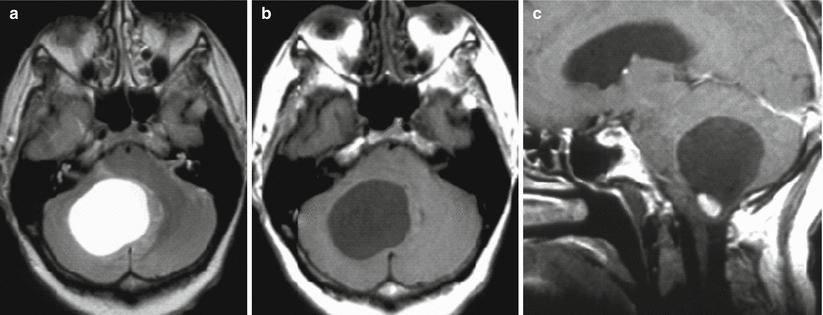

Fig. 33.1


Hemangioblastoma. On T2-weighted (a) and T1-weighted MRI (b), in the projection of the medial portions of the right cerebellar hemisphere, there is a cystic lesion with a mural nodule, which is located at the level of the foramen magnum and well visualized on T1-weighted MRI with contrast enhancement (c)
Stay updated, free articles. Join our Telegram channel

Full access? Get Clinical Tree


