, Valery Kornienko2 and Igor Pronin2
(1)
N.N. Blockhin Russian Cancer Research Center, Moscow, Russia
(2)
N.N. Burdenko National Scientific and Practical Center for Neurosurgery, Moscow, Russia
Toxoplasmosis is a parasitic disease caused by cysts of Toxoplasma gondii, contained in the raw meat. The infection in humans is possible not only as a result of consumption of such meat but also through animal’s excrements, raw milk, blood transfusion, organ transplant, the use of non-sterile needles, cat litter, as well as via an intrauterine route. The causative agent of the disease is widespread. From 6 to 90% of people in different regions have anti-Toxoplasma antibodies, with up to 30% of the world population being infested on average. Toxoplasma much more commonly affects the internal organs (brain, lungs) in patients with AIDS.
On CT without contrast enhancement, toxoplasmic encephalitis has characteristic manifestations in the form of multiple iso- or hypodense areas located predominantly in the basal ganglia (76 to 88%) and corticosubcortical layer. The disease can affect the posterior cranial fossa. Hemorrhages are very rare. Dimensions of lesions range from ≤1 to ≥3 cm. (Osborn 2004). Post-contrast CT images demonstrate annular or nodular opacification. Annular opacification with the central region with decreased density is a common manifestation of toxoplasmosis. The rim of contrast enhancement is usually thin and smooth, but thick and uneven rims are encountered in large lesions (Post et al. 1983). The ring accumulating the contrast medium corresponds to an area of an intense inflammatory reaction, and an edema surrounds the periphery of the ring.
MRI with and without contrast enhancement is more sensitive to both new and chronic lesions of toxoplasmic encephalitis than pre- and post-contrast CT. On T2-weighted MRI, active lesions have a variable MRI signal. They can be more hyperintense than the brain substance and indistinguishable from the high-intensity edema area. There may be an iso- or hypointense lesion in the center, surrounded by a high-intensity edema area. On T1-weighted MRI, the signal from the affected area can be isointense or hypointense (Figs. 40.1 and 40.2). After administration of gadolinium, there is its annular or nodular accumulation in the active area, well distinguished from the surrounding hypointense edema. Typical is the uneven accumulation of the contrast agent with formation of a local thickening along the contour or at the center of the lesion. At the same time, some fragments of the rest of the cyst surface can remain non-contrast enhanced. MRI and CT patterns of contrast enhancement are generally similar. Hemorrhages in toxoplasmosis are rare.
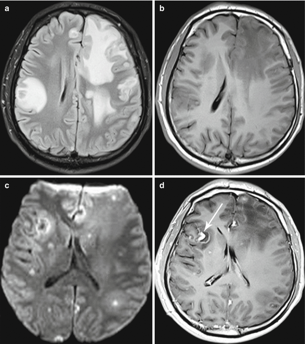
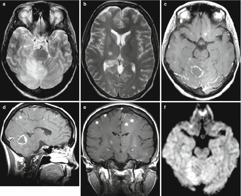
Fig. 40.1
Toxoplasmosis. On T2-FLAIR (a) and T1-weighted MRI (b), there are multiple foci with an increased MR signal in T2-FLAIR images and a decreased signal in T1-weighted images. On DWI MRI (c), there are multiple pinpoint areas with an increased signal; larger lesions are characterized by a heterogeneous signal. After CA administration, its intense accumulation in these areas is noted (d), with a clearly visualized, irregular thickening of the lateral wall in the largest lesion in the right frontotemporal area (the arrow)
Fig. 40.2


Toxoplasmosis. On T2-weighted (a, b) and T1-weighted MRI on the background of contrast enhancement (c–e), there are multiple foci of an increased MR signal and signs of intense accumulation of the contrast agent. On DWI (f), the signal is increased at the periphery of the largest lesion in the right hemisphere of the cerebellum
Related posts:


Stay updated, free articles. Join our Telegram channel
Full access? Get Clinical Tree
