11 Renal Artery
K.I. Ringe
The renal artery normally divides into two main trunks, which after branching in most cases into 5 to 7 arteries (range, 2–10) enter the parenchyma of the kidneys.1–5 They have no anastomoses within the kidneys and are “end arteries,” which supply individual segments or parts of segments. Polar arteries are therefore not auxiliary, supplementary, or accessory arteries as they have been called, for they replace the normal segmental branch. Polar arteries and multiple renal arteries are important in kidney transplantation.6
Renal segments have been included in the official anatomical nomenclature. Their functional relevance is debatable as the segments are based only on the arterial supply, but the arterial pattern can impact surgical approaches especially when nephron-sparing surgery is performed.7–13t
Atypical origins of the renal arteries are found more often in cases of ectopic or horseshoe-shaped kidneys but are also common in kidneys in normal positions. Ectopic origins of renal arteries have been described in individual cases from the following arteries: median sacral, external and internal iliac, lumbar, superior and inferior mesenteric, branches of the celiac trunk, and even cranial to the celiac trunk from the aorta.14–20
The high frequency of polar arteries, multiple arteries, and the origin of renal arteries from the aorta at different heights or from other arteries can be explained by the embryological development of the kidney. Many segmental arteries supply the pronephros, followed by the more cephalad mesonephros and finally the metanephros. There is a continuous involution of arteries and growth of new renal arteries. The persistence of some of these segmental arteries results in multiple renal arteries, polar arteries, or an abnormal origin of a renal artery.1,4,16,21–35
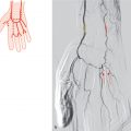
Although the right kidney has a more caudal position than the left kidney, the right renal artery originates, in general, more cranial to the aorta than the left renal artery.1,21,22,30,34,36 The right renal artery is longer than the left one because of the position of the aorta on the left of the vertebral column. The right renal artery usually runs posteriorly to the inferior vena cava and only anteriorly in about 4% of all cases when a single renal artery is present. In patients with two right renal arteries (in about 30%), one artery crosses the vena cava anteriorly.
There is considerable disagreement in the literature on the frequency of multiple renal arteries.37 This can, in part, be explained by the technical differences, for example, anatomical or corrosion cast preparation, aortographies, or selective renal arteriograms. In some patients with hypertension or hydronephrosis, a higher frequency of multiple renal arteries was described,38–42 although other studies showed no clear association between hypertension and accessory arteries.43 Multiple arteries sometimes occurred more frequently on the left side. The percentages given in the figures are the mean for both kidneys.
11.1 “Normal” Pattern as Described in Textbooks (59%)

Fig. 11.1 “Normal” pattern as described in textbooks (59%). Schematic of the right kidney (a) and coronal VR CT with normal branching patterns of the right and left renal arteries (b). In addition, marked atherosclerosis of the abdominal aorta can be appreciated.
11.2 Polar Arteries from the Renal Artery (15%)

Fig. 11.2 Upper polar artery from the main stem of the renal artery (13%). Schematic (a) and coronal MIP CT (b). 1 Upper polar artery; 2 renal artery.

Fig. 11.3 Lower polar artery from the main stem of the renal artery (2%). Schematic (a) and coronal MIP CT (b). Distinct atherosclerosis of the abdominal aorta is present. Status post stent implantation in the proximal main stem of the left renal artery (arrow). In addition, a complicated cyst in the lower pole of the left kidney can be appreciated (*). 1 Renal artery; 2 lower polar artery.

Fig. 11.4 Upper and lower polar arteries from the main stem of the renal artery (<1%). Schematic (a) and oblique coronal VR CT with upper and lower polar artery from the main stem of the renal artery (b). 1 Upper polar artery; 2 renal artery; 3 lower polar artery.
11.3 Two Renal Arteries (22%)

Fig. 11.5 Both renal arteries run to the hilus of the kidney (10%). Schematic (a) and CT (b,c). Oblique coronal VR (b) and coronal MIP (c) with two renal arteries on the right side. Lumbar arteries (*).
Related posts:


Stay updated, free articles. Join our Telegram channel

Full access? Get Clinical Tree


