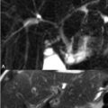
70 Renal Masses MRI of the normal kidneys clearly differentiates between the cortex and medulla, the latter exhibiting fluid-like SI due to the presence of urine. Cortical enhancement occurs at 20 to 30 seconds following contrast injection with uniform medullary and cortical enhancement at 80 seconds—the time at which masses are most sensitively detected. Simple renal cysts are the most common lesions in adults. A case of adult polycystic kidney disease is illustrated in Figs. 70.1A,B. Cysts of varying complexity are present, but the lesion denoted by the black arrow appears simple with typical low and high SI on (A) T1 and (B) T2WI, respectively, and no clearly identifiable wall or septation. The lesion denoted by the asterisk demonstrates SI compatible with a hemorrhagic cyst—high SI on (A) T1 and low SI on (B) T2WI—specifically the SI characteristics of intracellular methemoglobin. The vertebral body SI in the above figures is abnormal as these sequences were not acquired with fat saturation. The low SI correlates with the patient’s diagnosis of hemosiderosis (see Chapter 67). Simple cysts do not exhibit enhancement with contrast administration and are classified as Bosniak type I lesions. A Bosniak 2 cyst is illustrated in the (A) axial T2 and (B) contrast-enhanced T1WI of Figs. 70.2A,B, respectively. The (A) T2WI demonstrates a single, thin low SI septation that enhances minimally on (B
![]()
Stay updated, free articles. Join our Telegram channel


Full access? Get Clinical Tree


