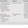
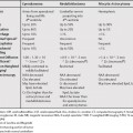
98 On T2-weighted magnetic resonance images (T2WI) the kidneys normally have moderately high signal intensity with no difference between the cortex and the medulla. Low signal intensity in the renal parenchyma could be secondary to hemosiderin, hemorrhage, calcification, or fibrosis. Three main categories of disease that result in renal parenchymal T2WI hypointensity are intravascular hemolysis, infection, and vascular disease. In extravascular hemolysis iron is deposited in the liver and spleen. In intravascular hemolysis, when direct release of hemosiderin in the plasma exceeds the binding capacity of plasma haptoglobin, hemoglobin is filtered in the glomerulus and stored in the proximal convoluted tubule as hemosiderin. Intravascular hemolysis does not result in hemosiderin accumulation outside the kidney.1,2 Intravascular hemolysis can be seen with mechanical shear (most commonly from a prosthetic valve), paroxysmal nocturnal hemoglobinuria, and rarely sickle cell anemia. Paroxysmal nocturnal hemoglobinuria is an acquired stem cell disorder caused by increased sensitivity to complement.1 Hemolysis, thrombosis, and deficient hematopoiesis are possible sequela.
Renal Parenchyma with Hypointensity on T2-Weighted Magnetic Resonance Imaging
Stay updated, free articles. Join our Telegram channel

Full access? Get Clinical Tree


