Abstract
Fetal renal pelvis dilatation is identified in 1% to 5% of pregnancies and is often a transient, normal variant. The likelihood of an underlying abnormality increases with the degree of dilatation. The most common underlying abnormalities are ureteropelvic junction obstruction and vesicoureteral reflux. Mild renal pelvis dilation is also a minor marker that confers slightly increased risk for trisomy 21. The pelvis is measured anterior to posterior in the transverse plane, with the calipers placed on the inner border of the fluid collection. Images in other planes (sagittal, coronal) are obtained to exclude evidence of obstruction elsewhere along the urinary tract. If the renal pelvis measurement exceeds a threshold in the second trimester (such as 4 mm before 20 weeks), further evaluation is recommended in the third trimester, at approximately 32 weeks. If there is calyceal dilatation or cortical thinning, more frequent evaluation may be warranted. Neonatal evaluation is typically reserved for those with renal pelvis dilatation of at least 7 mm in the third trimester.
Key Words
hydronephrosis, pyelectasis, calyceal dilatation, paradoxical hydramnios, ureteropelvic junction obstruction, vesicoureteral reflux
Introduction
Fetal renal pelvis dilatation (or dilation) is a common ultrasound (US) finding, identified in 1% to 5% of pregnancies. There is a strong correlation between the degree of renal pelvis dilatation and the likelihood of postnatal urinary pathology, particularly ureteropelvic junction (UPJ) obstruction and vesicoureteral reflux (VUR). However, the normal renal pelvis diameter also increases with advancing gestation, and mild dilatation is a transient, normal variant in most cases. The Society for Fetal Urology has termed fetal renal pelvis dilatation “a surrogate measurement of potential disease,” to denote that it does not represent pathology. Thresholds for renal pelvis dilatation in the second and third trimesters have been established to help in counseling families and guiding initial postnatal management ( Table 12.1 ).
| Degree of Dilatation | Second Trimester (mm) | Third Trimester (mm) |
|---|---|---|
| Mild | 4 to <7 | 7 to <9 |
| Moderate | 7 to ≤10 | 9 to ≤15 |
| Severe | >10 | >15 |
Conditions associated with renal pelvis dilatation as an isolated finding include UPJ obstruction and VUR. Renal pelvis dilatation may also occur in the setting of ureteral obstruction, as with duplication of the renal collecting system (see Chapter 13 ), and with urethral obstruction, such as posterior urethral valves (see Chapter 14 ).
Disorder
Definition
Fetal renal pelvis dilatation is also called urinary tract dilation or hydronephrosis , though these terms are less precise. It encompasses dilatation confined to the renal pelvis, termed pyelectasis or pelviectasis, and that which also involves the calyces, termed caliectasis or pelvicaliectasis . The pelvis is measured anterior to posterior in the transverse plane. Of the various thresholds used to diagnose dilatation, the most common are 4 mm in the second trimester (up to 20 weeks), and 7 mm at approximately 32 weeks in the third trimester. The second-trimester threshold is used to identify fetuses that need additional US evaluation in the third trimester, and it is also considered a minor marker that confers slightly increased risk for trisomy 21 (see Chapter 152 ). The third-trimester threshold is used to identify cases that need postnatal evaluation. Criteria have been established for mild, moderate, and severe renal pelvis dilatation, based on the risk of neonatal renal abnormality in a metaanalysis of more than 100,000 screened pregnancies. These definitions have been endorsed by the Society for Fetal Urology and are listed in Table 12.1 .
Prevalence and Epidemiology
Renal pelvis dilatation is observed in 1% to 5% of second-trimester pregnancies. The male-to-female ratio is 2:1. In 1.5% of pregnancies, approximately one-third of pregnancies with renal pelvis dilatation, a urinary abnormality is confirmed in the neonatal period. However, 40% to 90% of cases are transient and do not represent pathology. The overall likelihood of an underlying abnormality increases with the degree of dilatation: 12% with mild, 45% with moderate, and 88% with severe hydronephrosis.
The most common associated abnormality is UPJ obstruction, which occurs in 10% to 30% of cases. UPJ obstruction is identified in approximately 1:1000 to 1:2000 births, has a male-to-female ratio between 3:1 and 4:1, and is predominantly left-sided. The prevalence of UPJ obstruction increases with worsening dilatation, occurring in 5% with mild, 17% with moderate, and more than 50% with severe hydronephrosis. It is bilateral in 20% to 25% of cases.
VUR is also associated with renal pelvis dilatation. VUR occurs in approximately 4% with mild renal pelvis dilatation and 8% to 14% with moderate or severe pelvis dilatation. Although VUR is a less common cause of fetal hydronephrosis than UPJ obstruction, it is more frequent in the general population, occurring in approximately 1% of individuals. When VUR comes to attention in the neonatal period because renal pelvis dilatation was diagnosed prenatally, most affected infants are males, and VUR often resolves by early childhood. In contrast, when VUR is identified only after children have developed symptoms, the majority are females, and there is greater risk for renal insufficiency.
Etiology, Pathophysiology, and Embryology
The etiology of transient renal pelvis dilatation is unknown. It is postulated that minor kinks or folds in the ureter during development may lead to transient obstruction or reflux, or that the fetus may be experiencing ureteral relaxation from the effects of maternal progesterone. Mild renal pelvis dilatation up to 20 weeks of gestation is used as a minor marker that confers slightly increased risk for trisomy 21 (see Chapter 151 ).
The pathophysiology of UPJ obstruction is unknown and appears to be multifactorial. Potential causes include incomplete recanalization resulting in stricture, compression from fetal vessels, and abnormal innervation or muscular discontinuity that compromises normal peristalsis.
VUR is believed to occur when the ureteral bud develops at an abnormal location and makes contact with poorly differentiated portions of metanephric mesoderm, increasing the likelihood of hypoplasia or dysplasia. The location of the ureteral bud results in a vesicoureteral valve that is also abnormal, causing the reflux. VUR has been identified in up to 50% of parents and siblings of affected individuals, consistent with autosomal dominant inheritance.
Manifestations of Disease
Imaging Technique and Findings
Ultrasound.
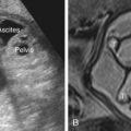
The fetal renal pelvis is measured anterior to posterior in the transverse plane, with calipers placed on the inner border of the fluid collection ( Fig. 12.1 ). Imaging should be performed in multiple planes, to identify the level of obstruction or any pathology along the course of the collecting system. In most cases, fetal renal pelvis dilatation is an isolated finding. The fetal ureter is not typically visible on US unless it is abnormal. Ureteral dilatation suggests reflux or obstruction at the level of the ureterovesical junction, as in the setting of a ureterocele (see Figs. 13.4 and 13.5 ). Marked hydronephrosis may also occur when there is obstruction at the level of the urethra, as with posterior urethral valves (see Fig. 14.6 ). A 2014 multidisciplinary consensus recommended that the sonographic description of renal pelvis dilatation include laterality, extent of calyceal dilatation, any parenchymal abnormality, bladder or urethral abnormalities, fetal sex, and assessment of amniotic fluid, and any abnormalities of other organ systems.

Related posts:


Stay updated, free articles. Join our Telegram channel

Full access? Get Clinical Tree


