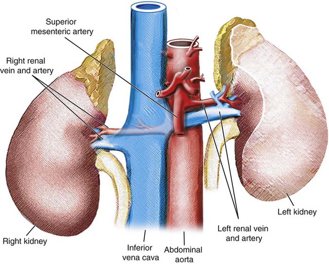
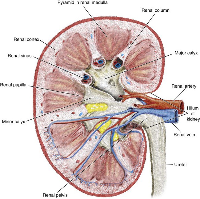
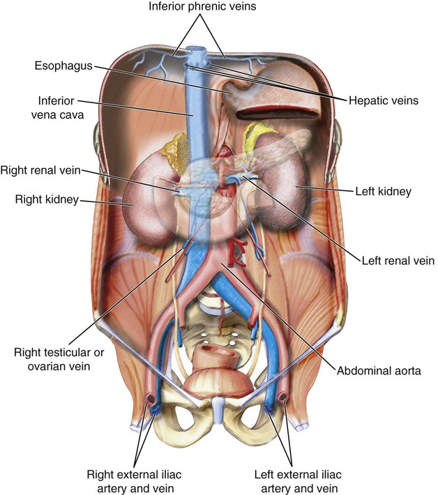
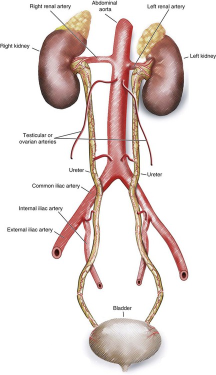
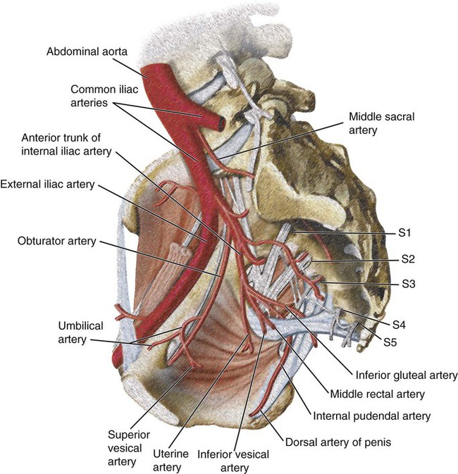
Renal Vasculature

Chapter 57


Renal Vasculature


Nadia J. Khati, Morvarid Alaghmand, Ali Noor, Aaron Reposar, Rishabh Chaudhari, Ramy Khalil and Anthony C. Venbrux


Introduction


Understanding the vascular anatomy of the kidneys, ureters, adrenals, and bladder is essential for image-guided therapy. For example, a patient with a traumatic fistula between the renal artery and vein may benefit from transcatheter embolotherapy. Such a procedure would spare the renal parenchyma and avoid emergent surgery.


Understanding the arterial blood supply to the ureters is important. In the clinical setting of reimplantation of the ureters into a neobladder, delayed distal ureteral strictures may develop secondary to ischemia. Such problematic lesions are due to disruption of the blood supply.


It is also important to have knowledge of the venous anatomy of the kidneys and adrenals. Sampling of renin levels from the renal veins may help determine a cause of hypertension (i.e., renovascular). Similarly, sampling from the adrenal veins may localize an aldosterone-producing tumor.


Renal Vascular Anatomy


Arterial Supply


The renal artery originates from the aorta at the area between L1 and L2, just inferior to the origin of the superior mesenteric artery (SMA). These arteries are typically posterior to the renal veins and anterior to the renal pelvis. The left renal artery arises at a higher level than the right renal artery and follows an upward course, but the right one is longer and passes posterior to the vena cava, with a downward course to the right kidney (Fig. 57-1).


As each renal artery approaches the hilum, it divides into anterior and posterior segmental arteries (Fig. 57-2). The posterior branch, which arises first, supplies a large portion of the posterior part of the kidney. Next, the four anterior divisions at the renal hilum arise as apical, upper, middle, and lower anterior segmental arteries. The apical and lower anterior divisions supply the anterior and posterior surfaces of the upper and lower renal poles. The upper and middle divisions supply the remaining parts of the anterior surface. The other divisions after the segmental arteries are the lobar, interlobar, arcuate, intralobular, and glomerular arteries. The renal artery gives off small branches to the renal capsule and adrenal gland (inferior adrenal artery).


Accessory renal arteries, which are the most common variant, can originate from the lateral aspect of the abdominal aorta, iliac, or renal artery (or rarely from the lower thoracic aorta or lumbar or mesenteric artery). They usually enter above or below the hilum. Perihilar arteries are other variations.


Of all documented renal vascular variants, accessory renal arteries are the most common and most clinically important. They are seen in up to one third of patients. Multiple accessory renal arteries are unilateral in 30% of patients and bilateral in approximately 10% of patients. Accessory arteries typically originate from the aorta or iliac arteries between the levels of T11 and L4 or rarely from the lower thoracic aorta, lumbar, or mesenteric arteries or a more proximal portion of the abdominal aorta above the origin of the SMA. Most accessory arteries flow into the renal hilum to perfuse both the upper and lower poles and are typically equal in size to a single renal artery. Less frequently, smaller accessory vessels are found to course directly into the renal parenchyma from the renal cortex and are thereby classified as polar arteries.


Perihilar arterial branching—branching of main renal arteries into branches at a point more proximal than the renal hilum—is also a common variant and especially important to note in preoperative mapping of renal transplant donors. In general, kidneys do not have efficient collateral circulation.


Venous System


As mentioned earlier, the renal veins are anterior to the renal arteries (see Fig. 57-1). The left renal vein is longer and crosses the midline anterior to the abdominal aorta and posterior to the SMA. This vein receives the left testicular or ovarian vein from below, the left suprarenal vein from above, and the lumbar vein before joining to the inferior vena cava (IVC; Fig. 57-3).


Ureteric Vasculature


As the ureters pass toward the bladder, they receive branches from adjacent vessels (Fig. 57-4). The renal arteries supply the upper end of the ureters, abdominal aorta, and testicular or ovarian arteries. The common iliac arteries supply the middle part, and the rest are supplied by branches from internal iliac arteries. There is anastomosis between these branches.


Dec 23, 2015 | Posted by in INTERVENTIONAL RADIOLOGY | Comments Off on Renal Vasculature

Full access? Get Clinical Tree

Get Clinical Tree app for offline access