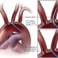
Restenosis is a multifactorial process involving three major mechanisms: remodeling, neointimal hyperplasia, and thrombosis. In vascular remodeling, arteries structurally adapt in size and composition. Using serial intravascular ultrasound imaging to observe the coronary artery after percutaneous transluminal coronary angioplasty (PTCA), Mintz et al.1 demonstrated that remodeling was a bidirectional phenomenon. Of 212 native coronary lesions in 209 patients after PTCA, 22% of the lesions showed positive remodeling (adaptive or expansive remodeling), and 78% of the lesions exhibited negative remodeling (constrictive or restrictive remodeling) with a decrease in lumen area (from 6.6 ± 2.5 mm2 to 4.0 ± 3.7 mm2; P < 0.0001). The SURE (Serial Ultrasound Restenosis) study2 examined the time course of this phenomenon by performing serial intravascular ultrasonography and revealed that the decrease in lumen and external elastic membrane volume occurred between 1 and 6 months after PTCA. The remodeling event was related to the adventitial fibrosis. Preliminary animal studies have demonstrated that arterial injury triggers the differentiation of adventitial fibroblasts into activated myofibroblasts by day 3 after injury.3 Myofibroblasts have both synthetic capability (type I collagen) and the ability to translocate to the vessel intima.4 They contribute to restenosis by proliferating, forming a fibrotic scar around the injury site, and migrating into the neointima.5 The source of the early proliferating cells causing stenosis formation is likely to be derived from the adventitia and media.6 Neointimal hyperplasia has been extensively investigated and described. First, the interventional damage to the endothelium and the subsequent exposure of subintimal collagen and tissue factors lead to loss of nitric oxide,7 prostacyclin,8 tissue plasminogen activator,9 heparan sulfate proteoglycans,10 and endothelial-derived hyperpolarizing factor.11 This process results in thrombosis, vasoconstriction, inflammation, and cellular activation and growth. Second, dysfunctional endothelial cells overexpress plasminogen activator inhibitor (PAI)-1, fibronectin, thrombospondin, integrins, selectins, angiotensinogen, angiotensin-converting enzyme, endothelin, and several growth factors, thereby leading to smooth muscle cell activation and growth.12 Proliferation plus migration of smooth muscle cells from the adventitial and medial layers of the vessel wall to the intima is a universal phenomenon in animal models of restenosis. However, the bulk of neointimal tissue is composed not primarily of smooth muscle cells themselves but of the fibrocollagenous extracellular matrix they secreted after activation. In human neointima, only 11% of the neointimal tissue mass is composed of cellular elements.13 The cytokines and growth factors released have the ability to promote proto-oncogene expression (e.g., c-myc, c-myb, c-fos), believed to regulate transformation of smooth muscle cells from a contractile phenotype to a secretory phenotype.14 The secretory phenotype of smooth muscle cells produces a loose extracellular matrix that represents the major portion of neointimal hyperplasia. Meanwhile, the whole process leads not only to tissue growth but also to fibrotic constriction of the adventitia, also responsible for constrictive remodeling.15 Mural thrombosis is a fundamental participant in the restenosis process. Immediately after vascular injury, circulating platelets adhere to exposed subintimal elements such as collagen, von Willebrand factor, fibronectin, and laminin via a number of glycoprotein membrane receptors. Platelet adhesion in turn promotes release of cytokines and growth factors, including platelet-derived growth factor, transforming growth factor (TGF)-β, and basic fibroblast growth factor, which can result in smooth muscle cell proliferation and, ultimately, neointima formation.16,17 Thrombus formation also acts as a scaffold for vascular smooth muscle cell migration, proliferation, and synthesis of matrix. The role of thrombosis in the restenosis process is supported by a clinical study by Bauters et al.18 In 117 consecutive patients who underwent successful PTCA and coronary angioscopy before/immediately after the procedure and at 6 months’ follow-up, they found that an angioscopically protruding thrombus at the PTCA site was associated with significantly greater loss of luminal diameter. Elastic recoil is a direct result of the elastic properties of the vessel wall, and it occurs immediately at the site of PTA. Ardissino et al.19 demonstrated that mean elastic recoil averaged 17.7% ± 16% in a study involving 98 consecutive patients and was correlated to the degree of residual stenosis immediately after coronary angioplasty (r = 0.64; P < 0.001). During 8 to 12 months of follow-up, no correlation was observed between the degree of elastic recoil and changes in minimal lesion diameter, but a positive correlation between the amount of elastic recoil and the incidence of restenosis was documented (r = 0.84; P < 0.05). Stenting virtually eliminates vessel elastic recoil and negative remodeling, the mechanical component of restenosis, but it also stimulates the cellular mechanisms that result in neointimal hyperplasia and subsequent ISR.20 In peripheral artery interventions, the iliac, femoropopliteal, and infrapopliteal arteries are distinguished by differences in interventional approaches and results.21 Primary patency of interventions is directly related to TransAtlantic Inter-Society Consensus (TASC) classification.22,23 In the iliac arteries, an endovascular approach is usually considered for TASC A and B lesions. Restenosis is not a principal limiting factor. The 2-year primary patency rate of iliac artery PTA/stenting was 81% to 83%.24,25 In the femoropopliteal region, restenosis is a major limitation. In a regression analysis of 330 consecutive patients undergoing femoropopliteal PTA or stenting, 6- and 12-month patency rates were 55% and 39% after PTA and 70% and 41% after stenting, respectively (P = 0.19).26 An analysis of 11 publications indicated that 1- and 3-year primary patency rates were 67% and 58% after femoropopliteal stenting.27 Recent studies showed that self-expandable nitinol stents appeared to perform better than PTA in the superficial femoral artery. The RESILIENT (Randomized Study Comparing the Edwards Self-Expanding LifeStent versus Angioplasty Alone in Lesions Involving the SFA and/or Proximal Popliteal Artery) trial showed 12-month freedom from target lesion revascularization was 87.3% for the stent group compared with 45.1% for the angioplasty group (P < 0.0001), and duplex ultrasound–derived primary patency at 12 months was better for the stent group (81.3% vs. 36.7%; P < 0.0001).28 It is reported that the 3-year patency rate of nitinol stents was as high as 76%.29 Infrapopliteal angioplasty has proved to be a reasonable primary treatment for critical limb ischemia (CLI) patients with TASC A, B, or C lesions. Freedom from restenosis, reintervention, or amputation was reported to be 39% at 1 year and 36% at 2 years.30,31 Higher patency rates can be expected with infrapopliteal stenting. In a randomized prospective study, 95 infrapopliteal lesions in 51 patients were treated by PTA or Carbofilm-coated stents. The 6-month primary patency rate of stenting was higher than that of PTA (45.6% vs. 79.7%; P < 0.05).32 Percutaneous transluminal renal angioplasty, alone or in conjunction with stenting, is a treatment strategy for atherosclerotic or fibromuscular dysplastic renal artery stenosis. In a prospective study of 31 fibromuscular dysplastic renal artery lesions in 27 patients, there was a cumulative 23% restenosis rate 12 months after PTA.33 Stenting has proved a better technique than PTA to achieve vessel patency in ostial atherosclerotic renal artery stenosis. In a nonrandomized prospective study of 163 patients with 200 atherosclerotic renal arterial lesions after PTA or stenting, there was a significant difference after PTA in 12-month patency rates of ostial, proximal, and truncal renal arterial stenoses (34% vs. 65% vs. 83%; P < 0.001), which essentially was based on a higher rate of restenoses in ostial lesions (P < 0.001). There was no statistically significant difference after stenting in patency rates of ostial, proximal, and truncal stenoses (80% vs. 72% vs. 66%). The stent-related reduction in relative risk of developing restenosis in 12 months was 70% in ostial stenoses (P = 0.002).34 In a randomized prospective study comparing PTA with stenting in 84 patients with ostial atherosclerotic renal artery stenosis, 6-month patency rates of PTA and stenting were 29% and 75%, respectively. Restenosis occurred in 48% of PTA and 14% of stenting procedures.35 The restenosis rate of renal artery stenting ranges from 15% to 20%.36 Stenoses of mesenteric arteries are usually focal and located at the ostium. Percutaneous endovascular PTA/stenting is becoming an alternative to surgery in patients with chronic mesenteric ischemia. The primary 1-year patency rates were 65% to 86%.37,38 In a retrospective analysis of 33 consecutive patients from a single institution who underwent PTA, stenting, or both for the treatment of chronic mesenteric ischemia, the primary long-term (mean, 38 months) clinical success rate was 83.3%, and the restenosis rate was 17.7%.39 Atherosclerosis of the brachiocephalic and subclavian arteries is not as common as that of the extracranial carotid arteries. The primary success rate was almost 100% for stenoses and 82% to 87% for total occlusions in the brachiocephalic and subclavian arteries. The 1-year primary patency rate was as high as 88% to 97.9%.40,41 In a study of 110 patients with symptomatic stenosis or occlusion of the proximal subclavian artery, PTA and stenting were technically and clinically successful in 102 patients. The 5-year primary clinical patency rate was 89%, with a median recurrent obstruction-free period of 23 months. The 5-year binary restenosis rate was as low as 13.7%.42 Carotid artery stenting has been proposed as an alternative to surgery for carotid artery obstruction in patients at high risk. In a meta-analysis of 34 studies on a total of 4185 patients with a follow-up of 3814 arteries over a median of 13 months, the cumulative restenosis rates after 1 and 2 years were approximately 6% and 7.5%.43 The patency of hemodialysis prosthetic grafts and arteriovenous fistulas is limited primarily by venous obstruction due to intimal hyperplasia and thrombosis. Stenosis can be treated by PTA, stenting, and in selected instances, atherectomy. Restenosis is still a principal factor in the low patency rate. PTA is the standard of care for treatment of vascular access stenosis and occlusions, providing a primary patency rate of 31% to 75% at 6 months.44 Life-table analysis revealed a 6-month patency rate of 63% and a 1-year patency rate of 41% for the first angioplasty in a given graft, and a 6-month patency rate of 44% and a 1-year patency rate of 22% for the second angioplasty.45 Systemic pharmacotherapies have targeted different mechanisms that have been identified as potential participants in the development of restenosis. Antithrombotic agents were associated with improved outcomes, especially cilostazol. In a study of 141 consecutive patients scheduled for PTA in the femoropopliteal artery, the incidence of TLR was significantly lower in the cilostazol-treated than in the non–cilostazol-treated patients (12% vs. 32%; P < 0.01).46
Restenosis
Background
Clinical Relevance
Peripheral Arteries
Renal Artery
Mesenteric Arteries
Subclavian and Carotid Arteries
Hemodialysis Access
Prevention of Restenosis
![]()
Stay updated, free articles. Join our Telegram channel

Full access? Get Clinical Tree


Radiology Key
Fastest Radiology Insight Engine


