A. Percutaneous biopsy
B. Whole-body PET-CT
C. Scrotal ultrasound
D. Metaiodobenzylguanidine (MIBG) study
2 A 62-year-old male presents with right groin pain. A hernia is suspected, and a CT of the abdomen and pelvis is performed. An incidental 5.1-cm fusiform infrarenal aortic aneurysm is identified. There are no comparison images. At what aneurysm sac size is repair indicated for an asymptomatic abdominal aortic aneurysm in an “average-risk” patient?
A. 4.0 cm
B. 4.5 cm
C. 5.0 cm
D. 5.5 cm
3 A 50-year-old male presents with an enlarged left external iliac lymph node. Percutaneous biopsy is planned. Which of the following approaches is least likely to result in a complication?
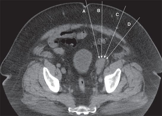
A. A
B. B
C. C
D. D
4 A 42-year-old male with pancreatitis and complicated peripancreatic fluid collections presents for catheter drainage. In which space is the fluid collection posterior to the left kidney located?
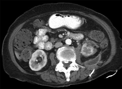
A. Anterior pararenal space
B. Posterior pararenal space
C. Perirenal space
D. Interfascial plane
E. Left paracolic gutter
5 A 60-year-old female with fatigue and no comorbidities undergoes a CT of the abdomen and pelvis demonstrating multiple enlarged retroperitoneal lymph nodes. A percutaneous core needle biopsy is performed under conscious sedation with a 15-cm-long 18-gauge end-firing biopsy device with a 2-cm throw. Six specimens are obtained, and the patient is taken to recovery.
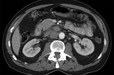
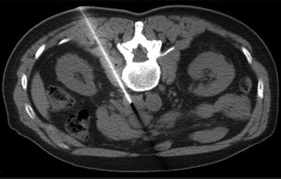
One hour later, the patient stands up for the first time since the biopsy, becomes queasy, and nearly loses consciousness. She is returned to her bed, and vital signs are obtained: (1) heart rate 40 bpm, (2) blood pressure 80/40 mm Hg, and (3) SpO2 100% on room air. She has no abdominal pain or ecchymosis. Two minutes later, she is asymptomatic, and her vital signs are (1) heart rate 72 bpm, (2) blood pressure 115/65 mm Hg, and (3) SpO2 100% on room air. What is the best next step?
A. Observation
B. Unenhanced CT
C. CT angiogram
D. Embolization
6 A 22-year-old male with metastatic testicular cancer undergoes a contrast-enhanced CT of the abdomen and pelvis. There is a single-comparison contrast-enhanced CT of the abdomen and pelvis performed 6 months ago. In addition to development of a new lesion, what defines “progressive disease” according to RECIST 1.1?
A. A ≥20% increase in the sum of diameters of the target lesions, with an absolute increase of at least 5 mm
B. A ≥30% increase in the sum of diameters of the target lesions, with an absolute increase of at least 5 mm
C. A ≥20% increase in the diameter of any single target lesion, with an absolute increase of at least 5 mm
D. A ≥30% increase in the diameter of any single target lesion, with an absolute increase of at least 5 mm
7 Which of the following structures is located in the anterior pararenal space?
A. Kidney
B. Duodenum
C. Transverse colon
D. Stomach
8 A 30-year-old female with right lower quadrant pain undergoes an abdominal MRI. Axial and sagittal T1-weighted contrast-enhanced GRE with fat saturation and axial and sagittal T2-weighted FSE sequences are presented below. What is the most likely diagnosis?
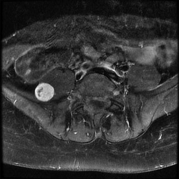
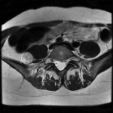
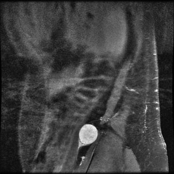
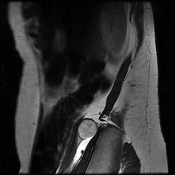
A. Paraganglioma
B. Nerve sheath tumor
C. Aneurysm
D. Metastasis
9 A 54-year-old male with stage V chronic kidney disease undergoes an unenhanced MR angiogram of his abdominal aorta. What best explains the “dark” appearance of fast-flowing blood on spin echo imaging?
A. Fast-flowing blood has a long T1.
B. Fast-flowing blood has a short T2.
C. Fast-flowing blood is only exposed to either a 90-degree or a 180-degree RF pulse.
D. Fast-flowing blood is always exposed to both a 90-degree and a 180-degree RF pulse.
10 A 25-year-old female stabbing victim undergoes a CT of the chest, abdomen, and pelvis. A 13-cm hemorrhage is identified in the right perirenal space related to a 3-cm renal laceration. Her hemoglobin is 7.1 g/dL. What best predicts this patient’s need for operative management?
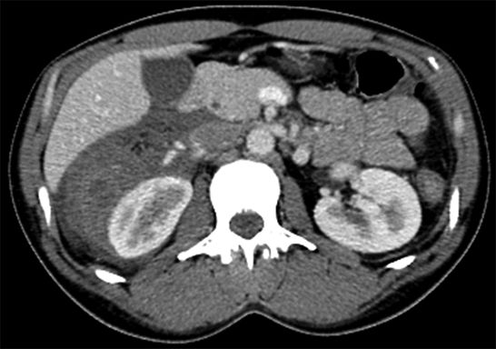
A. Mechanism of injury
B. AAST injury grade
C. Hemoglobin level
D. Size of the hematoma
E. Width of the laceration
11 A 51-year-old female with a 2.0-cm solid inferior pole right renal mass undergoes percutaneous radiofrequency ablation with two thermal ablation probes. An image from the ablation is shown below. What poses the greatest risk to this patient?
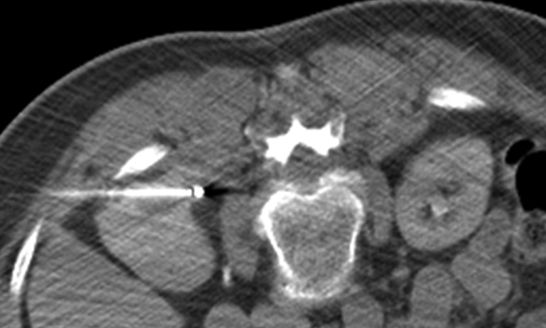
A. Nerve injury
B. Collecting system injury
C. Vascular injury
D. Incomplete treatment
12 A 65-year-old male with a remote history of colon cancer undergoes abdominal MRI after an incidental renal mass is discovered on surveillance CT imaging. Two unenhanced 2D axial gradient-recalled echo (GRE) images obtained from different imaging volumes are shown below. What is the most likely explanation for the appearance of the veins (left image) and arteries (right image)?
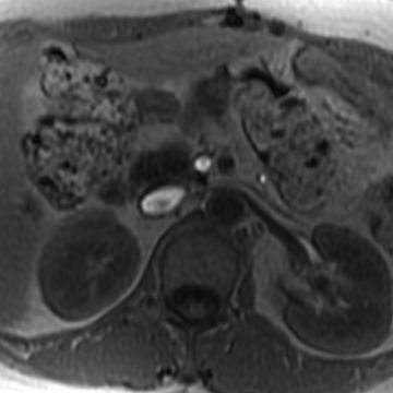
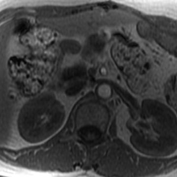
A. Unsaturated residual longitudinal magnetization entering the image volume
B. Unsaturated residual transverse magnetization entering the image volume
C. Saturated residual longitudinal magnetization entering the image volume
D. Saturated residual transverse magnetization entering the image volume
13 A 56-year-old asymptomatic male with microscopic hematuria undergoes a CT urogram that shows anterior displacement of the kidneys and duodenum. What is the best next step?
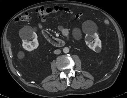
A. Clinical management (benign finding)
B. Percutaneous biopsy
C. Prophylactic ureteric stents
D. Operative debulking
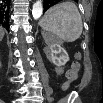
14 A 70-year-old male with dyspnea and long-standing pulmonary fibrosis undergoes a contrast-enhanced CT, and a lobular solid mass is identified. Representative images in the axial (left) and coronal (right) plane are shown below. What is the most likely diagnosis?
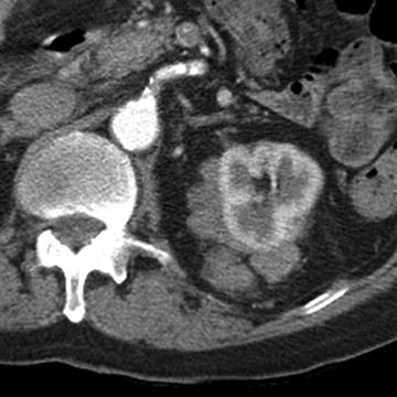
A. Renal cell carcinoma
B. B-cell lymphoma
C. Splenic angiosarcoma
D. Adrenocortical carcinoma
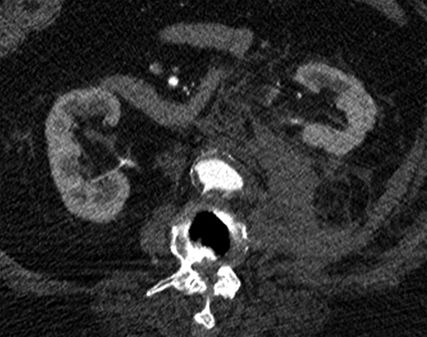
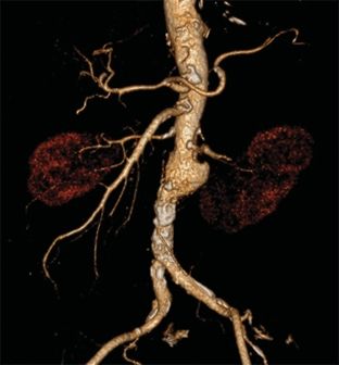
15 A 70-year-old male with a 40-pack-year smoking history and L2-L3 spondylodiscitis presents with acute flank pain, and a contrast-enhanced CT of the abdomen and pelvis is performed. The maximum infrarenal aortic caliber is 3.5 cm. What is the best next step?
A. Volume replacement and observation
B. Volume replacement and intravenous antibiotics
C. Volume replacement and emergent aortic endograft placement
D. Volume replacement and emergent operative repair
16 What are the typical findings of retroperitoneal Erdheim-Chester disease?
A. Diffusely enlarged sausage-shaped pancreas with a rind of peripancreatic fibrosis
B. Bilateral perirenal and pararenal soft tissue masses with associated ureteric obstruction
C. Unilateral locally aggressive solid mass of the renal capsule with or without calcifications
D. Bilateral renal and perirenal masses that displace but do not invade nearby structures
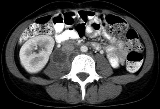
17 A 35-year-old female with fever and low back pain undergoes a contrast-enhanced CT of the abdomen and pelvis. The patient lives in the United States and has no relevant history. What is the most likely offending organism?
A. Staphylococcus aureus
B. Mycobacterium tuberculosis
C. Escherichia coli
D. Bacteroides species
18 A 70-year-old female with atrial fibrillation on warfarin therapy develops severe abdominal pain after a fall. What is the typical range of attenuation for clotted blood on an unenhanced CT of the abdomen?
A. 0 to 20 HU
B. 20 to 40 HU
C. 40 to 70 HU
D. 70 to 100 HU
19 A 60-year-old male with a 5.5-cm infrarenal aortic aneurysm undergoes percutaneous repair with an aortoiliac endograft. One year later, the sac size is 4.0 cm and a small type II endoleak is identified. What is the best next step?
A. Annual observation
B. Percutaneous embolization
C. Surgical ligation
D. Endograft excision
20 A 60-year-old male with vague low back pain undergoes a contrast-enhanced CT of the abdomen and pelvis. Which of the following diseases is associated with this imaging finding?
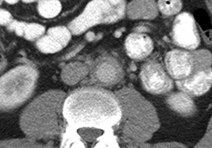
A. Renal cell carcinoma
B. Autoimmune pancreatitis
C. Autosomal dominant polycystic disease
D. Schistosoma haematobium infection
21 A 25-year-old female who is postoperative day 3 following an emergent cesarean section for fetal distress presents with sepsis and acute respiratory distress syndrome. An abdominal radiograph is performed. What is the most likely diagnosis?
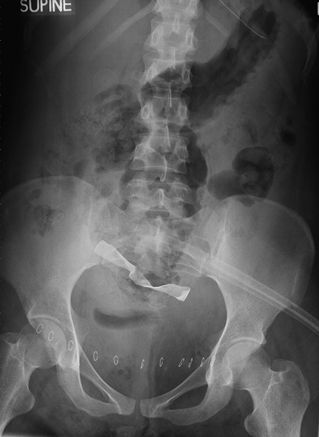
A. Necrotizing fasciitis
B. Large bowel obstruction
C. Retained foreign body
D. Malpositioned tube
22 A 54-year-old female who is postoperative day 9 following a partial right nephrectomy presents with persistent flank pain. Axial contrast-enhanced CT of the abdomen is performed (left), and coronal reconstructions are generated (right). What is the most likely diagnosis?
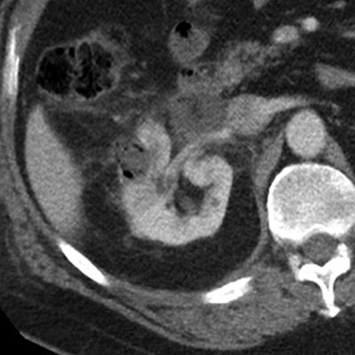
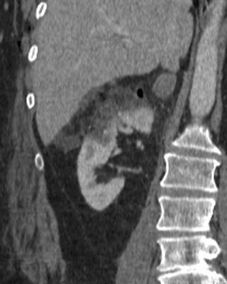
A. Normal postoperative appearance
B. Renal artery pseudoaneurysm
C. Collecting system injury
D. Renal abscess
23 Persistence of the right and left supracardinal veins can result in a “duplicated” or “double” IVC. When this variant manifests, into what structures do the right and left IVC usually drain?
A. Right IVC: right atrium; left IVC: azygous vein
B. Right IVC: azygous vein; left IVC: hemiazygous vein
C. Right IVC: right atrium; left IVC: left renal vein
D. Right IVC: right renal vein; left IVC: left renal vein
24 A 65-year-old male with stage V chronic kidney disease presents for an unenhanced MR angiogram. Conventional “black blood” spin echo sequences are planned. For a conventional spin echo–based sequence, when should the 180-degree refocusing pulse be applied?
A. Immediately before TE
B. Immediately after TE
C. TE/2
D. 2*TE
25 A 65-year-old male with Gleason 5 + 4 = 9 prostate cancer undergoes a contrast-enhanced CT of the abdomen and pelvis. What is the approximate sensitivity of morphologic CT imaging for the detection of nodal metastasis (on a node-by-node basis)?
A. 30%
B. 50%
C. 70%
D. 90%
26 A 68-year-old female undergoes a contrast-enhanced CT of the abdomen and pelvis that reveals a large retroperitoneal mass. What is the most likely cell type?
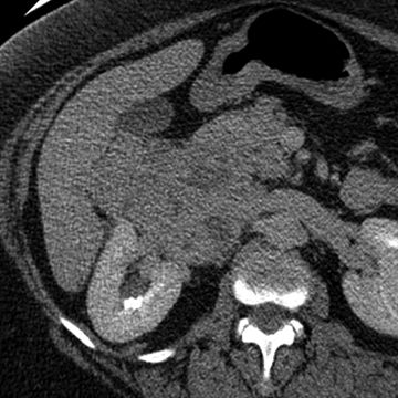
A. Liposarcoma
B. Leiomyosarcoma
C. Lymphoma
D. Leiomyoma
E. Lipoma
27 A 65-year-old male with lymphoma enrolls in a clinical trial and undergoes a contrast-enhanced CT reconstructed at 5-mm collimation. What criterion determines whether the lymph node depicted in the image would be considered a “target lesion” according to RECIST 1.1?
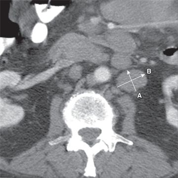
A. The node qualifies as a “target lesion” if line A is ≥10 mm.
B. The node qualifies as a “target lesion” if line A is ≥15 mm.
C. The node qualifies as a “target lesion” if line B is ≥10 mm.
D. The node qualifies as a “target lesion” if line B is ≥15 mm.
28 A 57-year-old female with adrenocortical carcinoma enrolls in a clinical trial and undergoes a contrast-enhanced CT reconstructed at 5-mm collimation. What criterion determines whether the mass depicted in the image would be considered a “target lesion” according to RECIST 1.1?
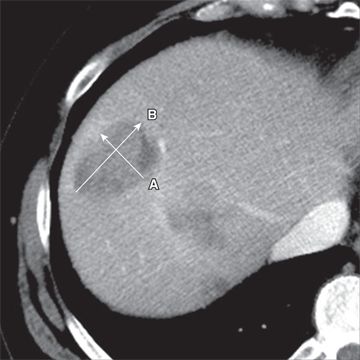
A. The mass qualifies as a “target lesion” if line A is ≥10 mm.
B. The mass qualifies as a “target lesion” if line A is ≥15 mm.
C. The mass qualifies as a “target lesion” if line B is ≥10 mm.
D. The mass qualifies as a “target lesion” if line B is ≥15 mm.
29 A 60-year-old male undergoes a CT angiogram. Isotropic images are acquired in the axial plane. Which of the following lines best represents how the aneurysm sac should be measured?
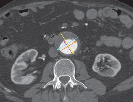
A. Blue
B. Yellow
C. Orange
D. Pink
E. The aneurysm sac should not be measured on this image.
30 A 60-year-old male with flank pain and hematuria undergoes a retroperitoneal ultrasound. A 4.5-cm solid renal mass is identified, and there is echogenic material in the renal vein and vena cava. Which of the following best distinguishes tumor thrombus from bland thrombus?
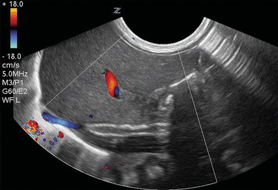
A. Tumor thrombus is echogenic and bland thrombus is hypoechoic.
B. Tumor thrombus has a systolic waveform and bland thrombus does not.
C. Tumor thrombus is central and bland thrombus is peripheral.
D. Tumor thrombus is mobile and bland thrombus is not.
Answers and Explanations
1 Answer C. The image demonstrates an enlarged low-attenuation interaortocaval lymph node near the insertion of the right gonadal vein. In a young male patient, isolated lymph node enlargement in this location is suggestive of metastatic testicular cancer. Of course, other etiologies are possible, and if the scrotal ultrasound is normal, the node would require further workup. Percutaneous biopsy (Answer A) is not the best first step because access to the node is challenging, and there are potentially noninvasive means of establishing the diagnosis (e.g., scrotal ultrasound). PET-CT (Answer B) is premature and probably unnecessary. An extra-adrenal paraganglioma, though possible, is less likely than a malignant lymph node, and therefore MIBG scan is not the best first step (Answer D).
Remember that in most patients, the right gonadal vein inserts upon the inferior vena cava just below the insertion of the right renal vein. The left gonadal vein inserts directly into the left renal vein. Nodal enlargement adjacent to either gonadal vein insertion site in an at-risk population is suggestive of testicular cancer, and scrotal ultrasound should be considered.
A generic differential diagnosis for an abnormal low-attenuation abdominal lymph node includes (1) germ cell tumor metastasis, (2) tuberculosis, (3) Whipple disease, (4) fungal infection, (5) necrotic metastasis, and (6) retroperitoneal lymphangioleiomyomatosis.
References: Pano B, Sebastia C, Bunesch L, et al. Pathways of lymphatic spread in male urogenital pelvic malignancies. Radiographics 2011;31:135–160.
Park JM, Charnsangavej C, Yoshimitsu K, et al. Pathways of nodal metastasis from pelvic tumors: CT demonstration. Radiographics 1994;14:1309–1321.
Stay updated, free articles. Join our Telegram channel

Full access? Get Clinical Tree


