CHAPTER 31 Rhabdomyosarcoma
Epidemiology
Rhabdomyosarcoma is the second most common pediatric head and neck malignancy and the most common soft tissue sarcoma in children. Rhabdomyosarcomas constitute 3% of all malignancies in patients younger than 20 years old.
Rhabdomyosarcomas are the most common primary temporal bone malignancy in children. These tumors most commonly present in children younger than 5 years old or in their late teens.
Approximately 7 to 8% of head and neck rhabdomyosarcomas involve the middle ear or mastoid regions (the nasopharynx and orbit are more common locations for these tumors).
In the middle ear and mastoid regions, these tumors are usually invasive and, as they are located near meninges, tend to spread intracranially. They are often unresectable at the time of diagnosis and have a relatively poorer prognosis than other head and neck rhabdomyosarcomas. However, less than 30% of patients have local metastases at presentation and there is an 85 to 90% three-year survival in these patients.
Clinical Features
Presenting symptoms are similar to those of chronic otitis media (which is a much more common diagnosis particularly in children), which is why the correct diagnosis is often delayed. Symptoms are similar to other middle ear processes including hearing loss, ear pain, ear discharge, and sometimes a bleeding external auditory canal (EAC) polyp. Facial nerve paralysis is unusual. The presence of an EAC polyp in a patient with chronic otitis should suggest the possibility of an underlying neoplasm.
Pathology
Rhabdomyosarcomas are highly malignant mesenchymal (skeletal muscle origin) tumors that are usually unresectable at presentation. There are three histologic types: embryonal (>75% of tumors), alveolar, and pleomorphic.
Treatment
Most tumors are not totally resectable at the time of diagnosis. Treatment usually entails debulking surgery (at which time the diagnosis is made) followed by multiagent chemotherapy and radiation therapy. Improvements in chemotherapeutic and radiation treatment protocols have resulted in up to 90% three-year survival in recent studies.
Imaging Findings
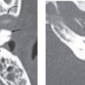
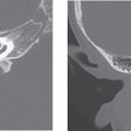
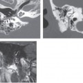
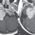
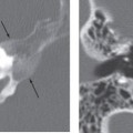
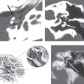
Like most malignancies of the temporal bone (and particularly the middle ear region), imaging is nonspecific and is performed to assess the extent of disease. Rhabdomyosarcomas of the temporal bone tend to be unresectable at presentation, so it is particularly important to define the extent of (extratemporal) spread of neoplasm before initiating therapy.
CT
Stay updated, free articles. Join our Telegram channel

Full access? Get Clinical Tree


