Rhabdomyosarcoma


Rhabdomyosarcoma


Shannon M. MacDonald

Alison M. Friedmann

Nancy J. Tarbell

Louis S. Constine


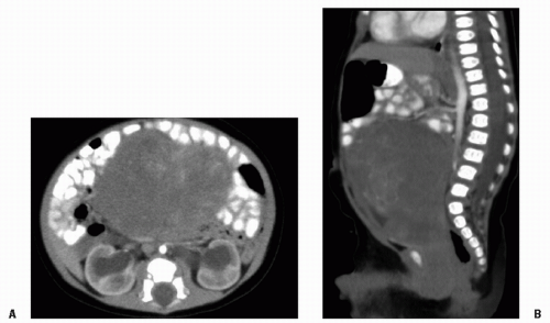
Rhabdomyosarcoma (RMS) is a highly malignant neoplasm that arises from embryonal mesenchyme with the potential for differentiating into striated muscle. Although the cells show differentiation along rhabdomyoblastic lines, RMS is not limited to cells with recognizable muscle cross-striations (1,2). Weber initially described RMS in 1854, but progress in our understanding of this complex neoplasm accelerated with Stout’s landmark descriptive series in 1946 and the delineation by Horn and Enterline in 1958 of the four classic forms of RMS (3, 4, 5). RMS can arise almost anywhere in the body, is locally invasive, and rapidly disseminates early in its course. Near the beginning of the 20th century, the only cures were accomplished with radical surgery in the few fortunate children without metastases. Significant disfigurement and loss of function were common sequelae. High-dose radiation therapy (RT) increased the potential for local control but caused a different set of morbidities (
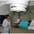
6,7). As chemotherapy has become increasingly effective in eliminating micrometastatic disease and assisting in local control, the need for aggressive surgery and large-volume irradiation has diminished, although surgery and RT still play pivotal roles in the curative treatment of RMS (8). Overall survival rates have concomitantly increased from 15-25% to more than 70% (9, 10, 11, 12).

RMS is a rare tumor with clinical and biologic heterogeneity. Consequently, multi-institutional trials were necessary to develop and refine treatment approaches. North American investigators of the Intergroup Rhabdomyosarcoma Study Group (IRSG) have played a paramount role in this progress, now in its sixth generation of protocols. The difficulty of this undertaking is clear in view of the myriad sites, stages, and histologies of RMS that are associated with different natural histories and prognoses (13). Beyond this, advances in imaging, changes in endpoints, and the need for mature data all increased the difficulty in conducting and comparing randomized, controlled clinical studies of patients with RMS. With this view, the IRSG was established through the collaboration of three multidisciplinary cancer treatment study groups (Cancer and Leukemia Group B, Children’s Cancer Study Group, and the Pediatric Branch of the Southwest Oncology Group, which later became the Pediatric Oncology Group). Intergroup Rhabdomyosarcoma Study I (IRS-I) was open for patient entry from 1972 to 1978 (9). With an overall 5-year survival rate of 55% in IRS-I, IRS-II was designed to improve survival for the patient subgroups with poor outcomes and to refine treatment for the remaining patients (10). IRS-II ran from 1978 to 1984, IRS-III from 1984 to 1991, IRS-IV from 1991 to 1997, and IRS-V from 1997 to 2005-2006. Although the previous generations of IRS studies were based on a surgically oriented clinical grouping system dependent on the tumor that remained after initial surgery, IRS-IV and IRS-V were based on a more biologically oriented staging system, discussed later in this chapter. These studies, each based on the results of its predecessor, have provided a database of over 4000 patients. The IRSG studies are now conducted through the Children’s Oncology Group, the United States-based cooperative research network that formed from the merger of the Pediatric Oncology Group and the Children’s Cancer Study Group. The current generation of studies opened in 2005-2006, upon the completion of IRS-V. Other multi-institutional group studies have also provided important data (14, 15, 16, 17). Of particular note are the Europeanbased International Society of Pediatric Oncology (SIOP) and the United Kingdom-based Children’s Solid Tumor Group (CSTG) (15, 16, 17). Finally, many single-institution studies have also been quite informative regarding RMS (7,8,18,19). Progress in treating RMS is exemplified by the improvement in overall 5-year survival in the IRS studies: 55% in IRS-I, 63% in IRS-II, and 71% in IRS-III (
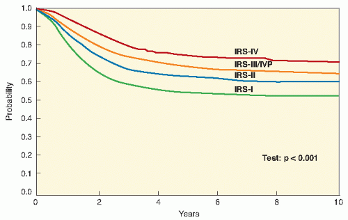
Fig. 11.1) (9, 10, 11,20). Early results from IRS-IV show an overall 3-year survival rate of 86% for patients with nonmetastatic disease and 39% for patients with metastatic disease (12,21).


EPIDEMIOLOGY

RMS accounts for about 3.5% of all malignant disease in children younger than 15 years of age and 2% of cancer cases among adolescents and young adults 15-19 years old (22,23). RMS is the most common soft tissue sarcoma of childhood, representing about half of this otherwise very heterogeneous group of tumors. The annual incidence of RMS is 4.4 per million in white children and 1.3 per million in black children. There is a slight male predominance (1.4:1). Seventy percent of cases occur before the age of 10 years, with a peak incidence at 2-5 years of age (Fig. 11.2). Congenital anomalies have been identified in as many as one third of children with RMS, most commonly involving the gastrointestinal, genitourinary, cardiovascular, and central nervous systems (24).

Although the majority of cases of RMS occur sporadically, a small proportion is associated with genetic conditions, including the Li-Fraumeni syndrome (in which germ line mutations of p53 exist), neurofibromatosis type 1, Costello
syndrome, Noonan syndrome, and Beckwith-Wiedemann syndrome (25, 26, 27, 28).


Figure 11.1 Improvement in 5-year survival of children with RMS treated on the IRS trials. (From Wexler L, Meyer WH, Helman LJ. Rhabdomyosarcoma and the undifferentiated sarcomas. In: Pizzo P, Poplack D, eds. Principles and Practice of Pediatric Oncology. 5th ed. Philadelphia, PA: Lippincott Williams & Wilkins; 2006:988, with permission.)


BIOLOGY

Although the origin and genetics of RMS remain unclear, clinical observations can provide direction for additional understanding of this tumor. If we can determine the tumor characteristics that predict radiochemotherapy responsiveness or, conversely, tumor resistance to therapy and a propensity to disseminate, then we can select the patients in whom more or less toxic therapy should be used. Moreover, we may devise novel biologic maneuvers to treat such tumors that minimize normal tissue damage. Reducing the toxicity of therapy becomes increasingly important as the cure rate continues to improve. Areas in which progress has been made include the genetic control of myogenesis, tumor suppressor genes, and molecular diagnostics (29,30).


Figure 11.2 Clinical features of RMS from IRS-I, IRS-II, and IRS-III pooled data. A: Age at presentation; (B) site of primary tumor; (C) clinical group; (D) histology. (Modified from Wexler L, Meyer, WH, Helman LJ. Rhabdomyosarcoma and the undifferentiated sarcomas. In: Pizzo P, Poplack D, eds. Principles and Practice Pediatric Oncology. Philadelphia, PA: Lippincott-Raven; 2006:976, with permission.)


Cytogenetics

Alveolar RMS is usually characterized by one of two translocations, both of which involve the FKHR gene on chromosome 13 (29,30). The most common translocation, t(2;13) (p35; q14), fuses the PAX3 gene, a transcription regulator, to the FKHR transcription factor. This translocation is present in about 60% of children with alveolar RMS. The less common translocation, t(1;13)(p36;q14), fuses the PAX7 transcription regulator to FKHR and is involved in about 20% of cases. This latter translocation seems to occur in younger patients and to be associated with a higher event-free survival rate than the PAX3 gene rearrangement. Approximately 20% of alveolar RMS does not have either translocation. The specific translocations (or lack thereof) do not appear to correlate well with histologic features (31). In the future, monitoring for these fusion products with sensitive techniques such as reverse transcriptase polymerase chain reaction may be useful for detecting minimal residual disease during or after treatment, and for guiding therapy (32,33).

Embryonal RMS often is characterized by loss of heterozygosity at 11p15.5, suggesting the presence of a tumor suppressor gene (34).

Genomic amplification also differs in alveolar and embryonal RMS. It is rare in embryonal RMS, whereas gains of whole chromosomes (particularly of chromosomes 2, 8, 12, and 13) and hyperdiploidy are common (35, 36, 37, 38). In alveolar RMS, gene amplification is common, as is near-tetraploidy.


Cell Cycle Control

Myogenesis involves differentiation of the mesenchymal fibroblast into skeletal muscle and is under the control of a series of gene products including the MyoD protein family (myogenin, MYF5, and MYF6) (39). These gene products also halt cell cycling. Expression of the MyoD proteins, which can be determined by an anti-MyoD antibody, has demonstrated that malignant cells have characteristics of skeletal muscle differentiation and therefore represent RMS. It is possible that some tumor suppressor factor is present in the normal fibroblast that, when combined with the RMS cell and the MyoD gene product, can drive the cell toward differentiation and halt cell proliferation.


Proto-Oncogenes

The myc gene can contribute to uncontrolled cell proliferation via several pathways including insertion mutations, translocations, retroviral transductions, and amplification. Two reports involving 20 patients showed that 50% of those with alveolar RMS had n-myc amplification, in contrast to its uniform absence in patients with embryonal RMS (40,41). Impressively, among the patients with alveolar RMS, n-myc amplification predicted a fatal outcome.


Tumor Suppressor Genes

Tumor protein p53 mutations can be demonstrated in a significant proportion of childhood RMS (42). In the presence of damaged DNA, normal p53 produces G1 arrest and prevents the cell with damaged DNA from undergoing any proliferation. In conjunction with myc, p53 drives such damaged cells toward apoptosis. This might explain why some forms of RMS, which escape cure by radiochemotherapy, become progressively more virulent: they generate resistant clones of tumor cells.


CLINICAL PRESENTATION

RMS occurs in any anatomic location where there is skeletal muscle and in some locations where skeletal muscle is not normally found (Fig. 11.2) (10). The most common locations are the genitourinary sites and the head and neck. Genitourinary sites include the bladder, prostate, vagina, uterus, urethra, and paratesticular region. Head and neck lesions are divided into parameningeal sites (nasopharynx, nasal cavity, paranasal sinuses, middle ear and mastoid region, infratemporal fossa, and pterygopalatine and parapharyngeal areas) and other head and neck sites (parotid region, cheek, masseter muscle, oral cavity, oropharynx, larynx, hypopharynx, scalp, face, and pinna) (43).

RMS commonly presents as a mass with poorly defined margins, but specific presentations relate to the primary disease site. A mass in the genitourinary system may cause urinary tract or rectal obstruction. On occasion, the mass may protrude from the cervix, vagina, or urethra. When the protruding tumor has the gross appearance of a cluster of grapes, it is called botryoid (grapelike) sarcoma. Prostate and bladder RMS may also have a botryoid appearance and protrude into the lumen of the bladder. Hematuria, urinary frequency, or retention with subsequent renal failure may occur. Paratesticular RMS presents with a mass and may be confused with a hydrocele, incarcerated hernia, or testicular torsion. In the extremities, RMS often is palpable and causes pain or limits motion. Parameningeal RMS may present with airway obstruction or a palpable mass. As the tumor grows, it can erode the base of the skull and cause cranial nerve palsies. Penetration into the brain can occur and mimic an intracranial mass, with headache, vomiting, and diplopia. RMS of the cheek or larynx causes obstruction of the aerodigestive track or a discernible mass; other symptoms or signs referable to the head and neck include hoarseness, polyps, decreased hearing, persistent otitis, otorrhea, rhinorrhea, nasal congestion or obstruction, and headache. Patients with orbital RMS usually present with proptosis, discoloration, or limitation of extraocular motion. RMS of the trunk can present as a mass simulating a hernia or hematoma, or causing a classic superior vena cava syndrome. In the retroperitoneum, RMS can cause gastrointestinal discomfort or other mass-related symptoms.


Diagnostic Evaluation

The history and physical examination should focus on the extent of local disease and the possible presence of metastases. RMS may extend locally and infiltrate along fascial planes and into surrounding tissues. Tumor margins often are indistinct. Depending on the site, the local tumor generally is imaged by some combination of computed tomography (CT), magnetic resonance imaging (MRI), and plain radiography. Genitourinary RMS often is investigated initially by ultrasound and barium enema, and voiding cystourethrogram, cystoscopy, or pelvic examination under anesthesia occasionally is indicated. The draining lymphatics, particularly in genitourinary and extremity primary sites, are evaluated with CT and often surgery
(44,45). The most common sites of metastases are lung, bone, bone marrow, and locoregional lymph nodes (21). Chest CT is the optimal imaging method for lung metastases. A nuclear medicine bone scan is performed to detect bony metastases but is not reliable in determining skull base involvement in parameningeal tumors, which is evaluated with CT or MRI. Bone marrow aspirate and biopsy are performed, and examination of cerebrospinal fluid (CSF) cytology is indicated if the tumor is in a parameningeal site (46). An MRI is used to evaluate spinal cord-related symptoms. Positron emission tomography (PET) scan is also being used with increasing frequency and is effective at detecting regional lymph node involvement and identifying distant metastases (47).


Table 11.1 International Classification of Rhabdomyosarcoma


Frequency (%)


Actuarial 5-Year Survival (%)


Superior prognosis


Botryoid rhabdomyosarcoma


6


95


Spindle cell rhabdomyosarcoma


3


88


Intermediate prognosis


Embryonal rhabdomyosarcoma


79


66


Poor prognosis


Alveolar rhabdomyosarcoma


32


54


Undifferentiated sarcoma


1


40


Other


9


Data from Newton WA Jr, Gehan EA, Webber BL, et al. Classification of rhabdomyosarcomas and related sarcomas. Pathologic aspects and proposal for a new classification: an Intergroup Rhabdomyosarcoma Study. Cancer. 1995;76:1073-1085.


Prognostic Features


Histology

Most RMS are soft, fleshy tumors with variation in the extent of both invasion and necrosis. Immunohistochemical stains, including antidesmin, antivimentin, and antimuscle-specific actin, are used routinely to help ascertain the muscle origin of the tumor cells, and the detection of the muscle regulatory gene MyoD1 may be even more sensitive than desmin (48).

Four histological subtypes of RMS are classically described: embryonal, alveolar, pleomorphic, and mixed (Fig. 11.2). However, the lack of agreement in classification among pathologists and the need to develop a single system that is prognostic prompted formation of an international panel to devise a new system, the International Classification of Rhabdomyosarcoma (Table 11.1) (49, 50, 51). Classification on the basis of histological features remains a challenge and current COG clinical trials utilize central pathologic review to ensure uniform pathological diagnosis since stratification schema and guidelines vary by histology (52).


Favorable: Embryonal, Botryoid, and Spindle Cell Variants

Embryonal RMS is the most common histological subtype. Embryonal RMS accounts for 60-70% of RMS in childhood and typically arises in the head and neck region and genitourinary tract (49). This form is composed of blastemal mesenchymal cells that tend to differentiate into cross-striated muscle cells. Although it resembles normally developing skeletal muscle in the 7-10 week fetus, great variation in the degree of differentiation can exist. Cellularity is moderate, and the stroma is loose and myxoid in most cases. The cells are generally fusiform or stellate, often admixed with primitive round cell forms. Cross-striations are present in about one third of cases (1). Periodic acid-Schiff staining, actin/desmin-positive reactivity, and Z-band material usually are present. Loss of heterozygosity at 11p15.5 may be identifiable. The pathologic differential diagnosis often includes lymphoma, Ewing’s sarcoma, and neuroblastoma (other small, round, blue cell tumors of childhood that have in common their light microscopic appearance and necessitate immunohistochemical evaluation for further characterization).

The botryoid variant of embryonal RMS represents about 10% of all RMS cases and occurs in mucosa-lined organs including the bladder, vagina, nasopharynx, nasal cavity, middle ear, and biliary tree. The stroma is loose with a myxoid character and a condensed tumor cell or cambial layer must be identifiable. The tumor cells may be small or large, with varying degrees of myogenesis. These tumors are generally localized and noninvasive (1,50,51).

The spindle cell variant is composed exclusively of spindleshaped cells and has a low cellularity. It can be collagen-rich or collagen-poor, with the former having a storiform pattern. Its most common site is paratesticular.


Unfavorable: Alveolar

About 20% of children with RMS have the alveolar subtype that is more common in adolescents and in tumors involving the extremities, trunk, and perianal and perineal region. The alveolar form resembles developing skeletal muscle in the 10-20 week-old fetus. The cells are round, with scanty eosinophilic cytoplasm that is occasionally vacuolated. Cross-striations are quite rare. The name alveolar is derived from the pattern produced by the tendency of cells to line connective tissue septa reminiscent of alveoli. Variable arrangement of trabeculae may cause the tumor cells to be arranged in strands, clefts, sheets, or clusters (1). The characteristic translocations are discussed in the “Biology” section earlier in this chapter. A “solid” variant has been identified, which grows as solid masses of closely aggregated cells with little or no discernible alveolar arrangement.


Table 11.2 Tumor, Node, Metastasis Pretreatment Staging Classification


Stage


Sites


Tumor Invasiveness


Tumor Size


Lymph Node Status


Metastasis


1


Orbit


Head and neck (excluding parameningeal)


Genitourinary (nonbladder, nonprostate)


Biliary tract


T1 or T2


A or b


Any N


M0


2


Bladder or prostate


Extremity


Head and neck parameningeal


Other (e.g., trunk, retroperitoneum)


T1 or T2


A


N0 or NX


M0


3


Same as stage 2


T1D or T2


A


B


N1


Any N


M0


4


All


T1 or T2


A or b


Any N


M1


T1, confined to anatomic site of origin; T2, extension; a, less than 5 cm in diameter; b, more than 5 cm in diameter; NX, clinical status unknown; N0, not clinically involved; N1, clinically involved; M0, no metastasis; M1, metastasis present.


Stage

RMS has been staged according to multiple systems developed in different institutions or multi-institutional groups. With the advent of IRS-III, a pretreatment staging system was developed based on the tumor, node, and metastasis (TNM) system used by SIOP, which reflected the disease characteristics at diagnosis (Table 11.2). IRS-IV, IRS-V, and the current COG trials have used this TNM staging system, which incorporates tumor size and invasiveness (a or b and T1 or T2, respectively), nodal status, presence of metastasis, and tumor site. Essentially, stage 1 tumors are in favorable sites. Stage 2 tumors are in unfavorable sites but are small (less than 5 cm) with negative lymph nodes. Stage 3 tumors are in unfavorable sites and are large or with positive lymph nodes. Stage 4 tumors are at any site, with hematogenous metastasis. This staging system has been validated with respect to its relationship to patient outcome (53).


Group

Implicit in the discussion of the grouping and staging systems is the importance of accurate identification of prognostic variables. Most of these variables are interrelated. A wealth of data supports the relevance of the clinical group of the patient, which in essence is the postsurgical disease extent at the time chemotherapy is initiated. The clinicopathologic grouping system has been used since the first IRS studies (Table 11.3, Fig. 11.2). The clinical group reflects the absence of microscopic disease (group I, 13% of patients) or presence of microscopic disease (group II, 20% of patients), gross disease (group III, 48% of patients), or metastatic disease (group IV, 18% of patients) (11,20). Clearly, the clinical group also reflects the disease site (ease of resection) and the biologic invasiveness of the tumor. Because therapeutic decisions made before study entry affected the assigned group, this system did not accurately reflect the biology of RMS (53, 54, 55). Moreover, the emphasis on surgical reduction of tumor bulk implicit in this system led surgeons to perform unnecessarily morbid surgeries at inappropriate times. In addition, the surgical approach was not uniformly applied, which obfuscated interpretation of results (54). However, data from all of the IRS studies support the utility of the grouping system, and it continues to be used in conjunction with stage and with careful surgical guidelines in the current trials that are specific to site of disease (Fig. 11.3) (9, 10, 11,20).


Primary Site

The primary site is a strong determinant of outcome, as verified by data from IRS-II and IRS-III (Fig. 11.4) (9, 10, 11,20). This relates, at least in part, to the association of site with other tumor and treatment variables. The primary site generally determines the ability to surgically remove the tumor, which in turn determines the IRS grouping (9). Surgical removal also relates to tumor invasiveness and the morbidity that would attend resection. Most orbital lesions are in group III (73.5% in IRS-I, IRS-II, and IRS-III); this is also true for parameningeal lesions (which are never in group I) and genitourinary bladder or prostate lesions (46,56). Conversely, most genitourinary nonbladder and nonprostate tumors are in group I, and most extremity tumors are in group I or II or are metastatic (group IV) at diagnosis. Other factors are also relevant to the association of primary site with prognosis. For example, the tumor location determines the presenting signs and symptoms, which are often related to the rapidity of diagnosis. Tumor size (up to 5 cm vs. more than 5 cm) is associated with survival time (p < 0.001) by multivariate analysis (51). Size is also related to tumor site through presenting symptoms and signs. Primary site influences the propensity for lymphatic spread (Table 11.4) (44,57). Whereas genitourinary, abdominal or pelvic, and extremity tumors commonly involve regional lymph nodes, tumors in the head and neck, trunk, and female genital organs rarely do so. However, the frequency of lymph node involvement is almost certainly underestimated by IRS data because the assessment of nodal status has not been systematic. Data from Stanford and Memorial Sloan-Kettering support the prognostic significance of lymph node involvement. Pedrick et al. (19) showed that 88% of patients presenting with involved nodes had primary tumors that were invasive and extended beyond the site or organ of origin. The more current IRS studies should provide more consistent documentation of lymph node status with improvements in imaging techniques and careful surgical guidelines.


Table 11.3 The IRS Grouping System


Group I


Localized disease, completely resected


a. Confined to muscle or organ of origin


b. Infiltration outside the muscle or organ of origin


Group II


Total gross resection with


a. Microscopic residual disease


b. Regional lymphatic spread, resected


c. Both


Group III


Incomplete resection with gross residual disease


a. After biopsy only


b. After major resection (more than 50%)


Group IV


Distant metastatic disease present at onset


Figure 11.3 Survival by clinical group for all patients treated in IRS-IV. Improved outcome was seen in group II compared with group I and presumably due to undertreatment of select patients with group I disease. (From Pizzo P, Poplack D, eds. Principles and Practice of Pediatric Oncology. Philadelphia, PA: Lippincott-Raven; 2006:984, with permission.)


Other Factors

A variety of other factors have prognostic significance, some of which are general and others specific to certain tumor subgroups. IRS data show that lymphocyte count, patient’s sex and age are prognostic (20). Although younger age is associated with a better outcome, the specific subgroup of children younger than 1 year old with alveolar histology have a significantly poorer survival than do older children, a finding not seen for infants with the embryonal subtype (58, 59, 60). Some site-specific variables influence outcome. In the head and neck, risk factors that predict tumor access to the cranial subarachnoid space (skull base erosion, cranial nerve palsy, intracranial extension) decrease the likelihood of disease-free survival (51% vs. 81% if no risk factors) (46,61,62). In extremity sites, the presence of lymph node involvement is strongly associated with a high incidence of relapse in metastatic sites and inferior survival. In contrast to many other childhood cancers, early response to chemotherapy has generally not been found to correlate with long-term disease-free control (63).


GENERAL PRINCIPLES OF THERAPY

Currently, the COG studies stratify patients into three risk groups (low, intermediate, and high) on the basis of known prognostic factors and historical outcome (13). The prognostic features incorporated into the risk classification scheme include histology, stage (which in turn incorporates site, tumor size, invasiveness, node status, and metastases), and clinical group. General definitions of the risk groups are as follows:


  • Low risk: Patients with localized embryonal RMS occurring at favorable sites (stage 1) and patients with embryonal RMS occurring at unfavorable sites with either completely resected disease (group I) or microscopic residual disease (group II)


  • Intermediate risk: Patients with embryonal RMS occurring at unfavorable sites with gross residual disease (group III) and patients with nonmetastatic alveolar RMS at any site


  • High risk: Patients with metastatic RMS (group IV, stage IV)

Clearly, this scheme will continue to evolve as treatments improve and new data emerge regarding particular subsets of patients and biologically based prognostic features. The overall goals are to reduce long-term toxicities in patients with a high likelihood of cure and to develop superior and innovative therapies for patients who continue to fare poorly with modern treatment regimens.

The therapeutic struggle for clinicians managing children with RMS, to cure while minimizing functional and cosmetic deficits, is heightened by the difficulty in eradicating both local and systemic disease. The spectrum of locations
and histologies complicates determination of treatment strategies through differences in the propensity for local and systemic control and treatment sequelae. It is clear that multidisciplinary therapy is necessary. Aggressive surgery and RT alone have been curative in fewer than 25% of children, with the exception of patients with orbital or genitourinary primary sites (17,64). Conversely, chemotherapy alone is associated with high local failure rates, a lesson learned by attempts to manage orbital or genitourinary tumors without radiation (17,65). The judicious use of chemotherapy to eradicate micrometastatic disease and reduce the extent of local disease and RT to increase the potential for local control has led to a decrease in aggressive surgery except in selected situations (8). The current challenge is to develop approaches to additionally enhance the complementary actions of all three treatment modalities in terms of intensity and sequence.


Figure 11.4 Event-free survival of patients treated on Intergroup Rhabdomyosarcoma Study IV by stage and site. A: For patients with nonmetastatic “favorable” site tumors (stage 1), the best outcome was seen for orbital primary tumors. B: For patients with nonmetastatic unfavorable site tumors (stage 2 or 3), the best outcome was for those with genitourinary (bladder-prostate) tumors, whereas those with extremity tumors had an inferior outcome. GU B/P, genitourinary tract (bladder or prostate); GU non-B/P, genitourinary tract (nonbladder, nonprostate). (From Pizzo P, Poplack D, eds. Principles and Practice of Pediatric Oncology. Philadelphia, PA: Lippincott-Raven; 2006:991, with permission.)


Surgery

Before the advent of radiation and chemotherapy, complete resection of RMS was the clear goal of surgical treatment. This often involved radical surgeries such as pelvic exenteration, radical prostatectomy, cystectomy, amputations, and orbital exenteration. Even so, fewer than 10% of children were amenable to complete resection and curable because of the absence of metastatic disease. Beyond this, most of these children had severely compromised quality of life functionally, cosmetically, and psychologically. Select sites that were more often curable by aggressive surgery included the orbit and bladder. Combined analysis of patients with nonmetastatic disease from IRS-I, IRS-II, and IRS-III showed that only 16% of children had disease amenable to complete resection (group I) and 20% had tumors for which removal of gross disease (group II) was possible (Fig. 11.5). In general, achievement of

local control with organ preservation is the appropriate goal. This is accomplished with appropriate combined-modality therapy. Aggressive surgery remains appropriate in certain situations, particularly for salvage therapy.


Table 11.4 Lymph Node Metastasis by Primary Site for 592 Patients with Visibly Resected Diseasea from IRS-I and IRS-II


Site


Number of Patients


Number and Percentage with Nodal Metastases


Extremity


Upper


74


12 (16%)


Lower


107


10 (9%)


Total


181


22 (12%)


Genitourinary organs


Paratesticular


107


28 (26%)


p = 0.001b


Bladder


29


6 (21%)


Prostate


12


5 (42%)


p = 0.03


Female genital organs


17


1 (6%)


Other


1


N


Total


166


40 (24%)


Head and neck


Orbit


39


0 (0%)


Other


96


8 (8%)


p = 0.06


Total


135


8 (6%)


Other


Anus and perineum


15


2 (13%)


Pelvis and retroperitoneum


22


5 (23%)


Trunk


65


2 (3%)


p = 0.01


Abdomen and thorax


8


2 (25%)


Total


110


11 (10%)


Totals


592


81 (14%)


aMicroscopic or no residual disease.

b p values relate to comparison of frequency of nodal metastases for this site compared with the 14% for all 592 patients. From Lawrence W Jr, Hays DM, Heyn R, et al. Lymphatic metastases with childhood rhabdomyosarcoma. Cancer. 1987;60:910-915, with permission.


Figure 11.5 Axial (A) and sagittal (B) images of a 10-month-old boy with a group III rhabdomyosarcoma of the bladder/prostate. The tumor displaces the bowel and causes urinary obstruction resulting in hydronephrosis.

Reoperation for microscopic residual disease after an initial excision, or when the first operation was performed without knowledge of the type of neoplasm involved, may be indicated before additional management. Reoperation after chemotherapy as a second-look procedure provides an attractive option for select cases. Many of these patients have had a “pathologic” complete response and have survival similar to that of patients who had an initial complete resection.

The role of lymph node dissection as a component of surgical therapy continues to evolve. Advances in imaging may improve radiographic detection of involved lymph nodes. Current guidelines are site specific because of the variability in the frequency of lymph node involvement and outcome data relating to its significance. Genitourinary and extremity RMS have a high incidence of lymph node metastases (Table 11.4) (44). The high frequency of nodal spread for extremity tumors, often without clinical or radiographic evidence of involvement, has both prognostic and therapeutic implications (66,67). Currently, COG guidelines include surgical evaluation of lymph nodes for RMS of the extremity and for males who are older than 10 years of age with paratesticular tumors. Sentinel lymph node biopsy continues to be explored as an alternative to formal lymph node dissection (68). This topic is considered further in later sections because considerations are site specific.

Overall, the extent and timing of surgical excision depend on the site of tumor and overall treatment strategy, balancing cure with functional outcome.


Chemotherapy

Before the 1960s, the role of chemotherapy in RMS was for the treatment of metastatic disease. Several investigators reported responses to vincristine and actinomycin used alone (VA) or in combination with cyclophosphamide (VAC) (64). Various groups then began to report that the adjuvant administration of chemotherapy for totally or subtotally resected localized disease contributed to an increase in survival probability from 10-40% to 60-80% (69). Heyn et al. (8) randomized 32 children with completely resected RMS to adjuvant therapy with VA or no adjuvant therapy. There were 8 deaths among the 15 children in the control group and 2 deaths in the 17 treated children. All children with microscopic residual disease received chemotherapy, and survival rates were excellent.


Table 11.5 Design and Results of IRS-I, 1972-1978 (686 Patients)


Clinical Group


Chemotherapy Regimen


Conventional Radiation Therapy


5-Year Survival (%)


Entire Group Survival (%)


I


VAC × 2 years


No


93


83


VAC × 2 years


Yes


81


II


Cyclic sequential VA × 1 year


Yes


73


71


VAC × 2 years


Yes


70


III


Pulse VAC × 2 years


Yes


53


52


Pulse VAC + Adr × 2 years


Yes


51


IV


Pulse VAC × 2 years


Yes


14


21


Pulse VAC + Adr × 2 years


Yes


26


Overall


55


A, actinomycin D; Adr, doxorubicin; C, cyclophosphamide; V, vincristine.


Modified from Mandell LR. Ongoing progress in the treatment of childhood rhabdomyosarcoma. Oncology. 1993;7:71-83, with permission.


With the widespread adoption of chemotherapy as part of the therapy of RMS, several trials were undertaken to establish the optimum drug combinations. IRS-I tested whether VAC was superior to VA in group II disease and whether pulse VAC plus adriamycin was superior to pulse VAC alone in groups II and III (Table 11.5). The study found no benefit to cyclophosphamide in group II diseases, and no benefit to the addition of adriamycin in groups III and IV (9).

IRS-II built on the results of IRS-I (Table 11.6) (10). Patients in group I received VAC or VA. Disease-free survival was similar. IRS-II showed no benefit to pulse VAC compared with cyclic sequential VA for group II. In groups III and IV, pulse VAC was better than a VAC and adriamycin combination but not statistically significantly better.

IRS-III (1984-1991) separated patients by histology into either a favorable (embryonal) or unfavorable (alveolar, anaplastic, and monomorphous) histology. Results based on subgroup are detailed in Table 11.7. Several drug pairs (adriamycin with imidazole carboxamide [DTIC], actinomycin D with VP-16, and actinomycin D with DTIC) appear to have been associated with gain in survival (11).

IRS-IV (1991-1997) used the new staging system to assign drug therapy. Patients with group I paratesticular tumors and group I or II orbital tumors were treated with VA, and all other nonmetastatic patients except those with preexisting renal abnormalities were randomized to receive one of three chemotherapy regimens: VAC; vincristine, actinomycin D, and ifosfamide (VAI); or vincristine, ifosfamide, and etoposide (VIE) (12). Patients with group III tumors were also randomized to receive conventional RT or hyperfractionated RT. VAC, VAI, and VIE were equally effective, and overall the patients with embryonal tumors who received three-drug therapy benefited in comparison to those in IRS-III, where just VA was given. For patients with metastatic tumors, a pilot IRS study looked at the activity of ifosfamide and doxorubicin in
an up-front window and found a response rate of 63% (complete and partial responses) (70). Subsequently, a full randomized IRS trial in 128 patients compared two up-front treatment windows, vincristine and melphalan (VM) and ifosfamide and etoposide (IE), and later therapy consisted of VAC or VAC with the window therapy included if the patient had an initial response (71). Initial response rates were comparable (VM 74% vs. IE 79%), but 3-year failure-free survival (FFS) and overall survival (OS) rates were superior in the patients who received the IE-containing regimen (FFS 33% vs. 19% and OS 55% vs. 27%).


Table 11.6 Design and Results of IRS-II, 1978-1984 (1003 Patients)


Clinical Group


Chemotherapy


Conventional Radiation Therapy


5-Year Survival (%)


Entire Group Survival (%)


Ia


VA × 1 year


No


85


81b


VAC × 2 year


No


84


IIa


Cyclic sequential VA × 1 year


Yes


88


80b


Repetitive pulse VAC × 1 year


Yes


79


III


Repetitive pulse VAC × 2 years


Yes


66


65


Repetitive pulse VAdrC-VAC × 2 years


Yes


65


IV


Repetitive pulse VAC × 2 years


Yes


26


27


Repetitive pulse VAdrC-VAC × 2 years


Yes


27


Overall


62


63


A, actinomycin D; Adr, doxorubicin; C, cyclophosphamide; V, vincristine.


a Patients with alveolar and extremity tumors excluded in groups I and II; treated with repetitive pulse vincristine, actinomycin D, and cyclophosphamide × 1-2 years with or without conventional radiotherapy.

b Includes extremity and alveolar tumors treated differently.


Modified from Mandell LR. Ongoing progress in the treatment of childhood rhabdomyosarcoma. Oncology. 1993;7:71-83; and


Maurer HM, Gehan EA, Beltangady M, et al. The Intergroup Rhabdomyosarcoma Study II. Cancer. 1993;71:1904-1922, with permission.

Only gold members can continue reading. Log In or Register to continue

Stay updated, free articles. Join our Telegram channel

Jun 19, 2016 | Posted by in GENERAL RADIOLOGY | Comments Off on Rhabdomyosarcoma

Full access? Get Clinical Tree

Get Clinical Tree app for offline access