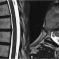
33 Rheumatoid and Postoperative Changes Rheumatoid arthritis exhibits a predilection for the cervical spine at the atlas and dens. Osteophytes are characteristically absent. Transverse ligament laxity and eventual atlantoaxial subluxation may occur. Such laxity is manifest as increased atlantoodontoid distance. Settling of the skull on the atlas is a severe complication and is demonstrated in Fig. 33.1A. In basilar invagination, by definition, the dens extends above Chamberlain’s line (drawn from the posterior hard palate to the posterior lip of the foramen magnum) by >3 mm. This case also demonstrates a large, hypervascular pannus surrounding the dens (in particular anteriorly) and exhibiting high SI on (A) T2WI. A hypervascular pannus will brightly enhance, although minimally enhancing hypovascular and fibrous panni also occur, demonstrating intermediate and low SI on T2WI, respectively. Erosion of the dens is clearly seen on the (B) axial T1WI. Although such erosions may be better visualized on CT, MRI offers superior soft tissue evaluation, such as the rheumatoid-related cord compression in Fig. 33.1A. Sagittal images in flexion and extension may further aid in evaluation. Ischemic cord damage resulting from chronic compression eventually warrants surgery to prevent further injury. An anterior approach is most common, whereby the disk is resected and replaced with bone graft. As seen with the anterior plate and screw fusion depicted on the sagittal T2WI image of Fig. 33.2A
![]()
Stay updated, free articles. Join our Telegram channel


Full access? Get Clinical Tree


