Presentation and Presenting Images
( ▶ Fig. 78.1, ▶ Fig. 78.2, ▶ Fig. 78.3, ▶ Fig. 78.4, ▶ Fig. 78.5)
A 35-year-old female with a palpable finding in the axillary tail of the right breast and left breast pain presents for diagnostic mammography.
78.2 Key Images
( ▶ Fig. 78.6, ▶ Fig. 78.7, ▶ Fig. 78.8, ▶ Fig. 78.9, ▶ Fig. 78.10)
78.2.1 Breast Tissue Density
The breasts are heterogeneously dense, which may obscure small masses.
78.2.2 Imaging Findings
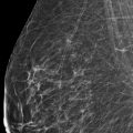
There was no mammographic finding corresponding to the palpable finding reported in the right axillary tail that is noted by the triangular skin marker (arrow in ▶ Fig. 78.6 and ▶ Fig. 78.8). The left breast demonstrates a 5-cm well-circumscribed, fat-containing mass central in the breast and located 3 cm from the nipple ( ▶ Fig. 78.7 and ▶ Fig. 78.9). Only the craniocaudal (CC) digital breast tomosynthesis (DBT) movie was done and demonstrates the finding it best on slice 26 of 83 ( ▶ Fig. 78.10).
78.3 BI-RADS Classification and Action
Category 0: Mammography: Incomplete. Need additional imaging evaluation and/or prior mammograms for comparison.
78.4 Diagnostic Images
( ▶ Fig. 78.11, ▶ Fig. 78.12, ▶ Fig. 78.13, ▶ Fig. 78.14)
78.4.1 Imaging Findings
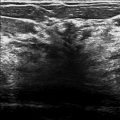
Ultrasound showed a mixed echoic encapsulated mass corresponding to the left breast mammographic finding ( ▶ Fig. 78.11 and ▶ Fig. 78.12). There was no sonographic correlate for the right axillary tail palpable finding (not shown).
The palpable finding was of great clinical concern so magnetic resource imaging (MRI) was also performed. The MRI showed no correlate for the right axillary tail palpable finding (not shown). Normal tissue was seen and clinical correlation was recommended. The mammographic and sonographic mass seen centrally in the left breast corresponds to an encapsulated mass containing both fat and breast tissue on MR ( ▶ Fig. 78.13 and ▶ Fig. 78.14). It has the classic appearance of a “breast within a breast.”
78.5 BI-RADS Classification and Action
Category 2: Benign
78.6 Differential Diagnosis
Hamartoma: The appearance of hamartomas depends on the fat to parenchyma ratio. Lesions with a greater amount of fat can mimic lipomas, those with a greater amount of parenchyma can mimic fibroadenomas.
Galactocele: The appearance depends upon the amount of fat and proteinaceous material and the viscosity of the fluid. Galactoceles are most commonly seen after the cessation of breast-feeding.
Lipoma: A lipoma usually presents on mammography as a well-circumscribed uniformly radiolucent mass.
78.7 Essential Facts
Hamartomas were first described as lipofibroadenomas, fibroadenolipomas or adenolipomas, based on the predominant component of the mass.
Hamartomas of the breast are uncommon masses with a reported incidence of 0.7% of all benign breast masses in females.
On physical examination, hamartomas may be occult or may present as mobile, soft-to-firm palpable masses.
Although not microscopically encapsulated, hamartomas have the appearance of a capsule or pseudocapsule on mammography and DBT.
Hamartomas have been described as a “breast within a breast” because mammographically they are commonly seen as a well-circumscribed round to oval mass surrounded by a thin capsule and composed of fat and breast tissue.
78.8 Management and Digital Breast Tomosynthesis Principles
DBT greatly reduces the effect of overlapping breast tissue, which results in the better visualization of mass margins than on conventional mammography.
Fat, even when not appreciated on conventional mammography, is seen in benign and malignant masses with DBT.
The greater detail seen with DBT imaging allows for definitive classification of encapsulated fat-containing masses.
The majority of encapsulated fat-containing masses found on mammography and DBT are benign.
78.9 Further Reading
[1] Arrigoni MG, Dockerty MB, Judd ES. The identification and treatment of mammary hamartoma. Surg Gynecol Obstet. 1971; 133(4): 577‐582 PubMed
[2] Freer PE, Wang JL, Rafferty EA. Digital breast tomosynthesis in the analysis of fat-containing lesions. Radiographics. 2014; 34(2): 343‐358 PubMed

Fig. 78.1 Right craniocaudal (RCC) mammogram.
Stay updated, free articles. Join our Telegram channel

Full access? Get Clinical Tree


