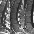
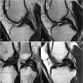
82 Rotator Cuff Tears A shoulder MRI may require initial acquisition of thin-section (≤3 mm) axial images with the shoulder in neutral or slight external rotation to properly position parasagittal and paracoronal slices parallel to the surface of the glenoid cavity and supraspinatus tendon, respectively. A dedicated extremity coil allows maximization of SNR and spatial resolution. Evaluation of the long head biceps tendon, neurovascular bundles, and the relationship between the humeral head and glenoid labrum is best performed in the axial plane, whereas the paracoronal slices (Figs. 82.1A,B,C,D) better demonstrate the rotator cuff muscles, tendons, and associated bursae. FSE T2WI (with or without fat suppression) must be acquired in all imaging planes for accurate characterization of pathology. Axial GRE T2WI and MR arthrography are useful for evaluation of the glenoid labrum. Hyaline cartilage lining the articular surfaces of the humeral head and glenoid cavity normally demonstrates moderate SI on T1WI and T2WI, whereas fibrocartilage, which lacks free mobile protons, composes the glenoid labrum and articular surface of the acromioclavicular (AC) joint and appears of low SI on conventional pulse sequences. Fibrous rotator cuff tendons similarly demonstrate low SI, impairing their differentiation from adjacent low SI cortical bone. Tendinosis, partial tears, and small full-thickness tears of the rotator cuff represent a continuum of disease and may be difficult to distinguish. Tendinosis demonstrates increased SI on T1WI or PDWI. SI on FS T2WI is less than that of fluid, as illustrated in the case of supraspinatus tendinosis in Fig. 82.1A
![]()
Stay updated, free articles. Join our Telegram channel

Full access? Get Clinical Tree


