Scoliosis


Scoliosis


Lindsay J. Rowe

Terry R. Yochum

Chad J. Maola

Norman W. Kettner


GENERAL CONSIDERATIONS

The term scoliosis is usually credited to Hippocrates. (1) Its derivation is from the Greek word skolios, meaning “twisted” or “crooked.” In general, any lateral curvature of the spine > 10° in the coronal plane is called a scoliosis, though many use the term to describe any lateral spinal deviation. Curves of 5-10° have been found in up to 15% of school pupils. (2) The incidence of scolioses > 10° is estimated between 2% and 4% in the general population. (3) The common right (dextro) thoracic curve, measuring < 10°, is generally considered physiologic and is thought to accommodate the size and position of the heart, lung, and aorta or to be related to handedness. (4) Spontaneous resolution or improvement of curves < 10° can be observed in 3-20% of individuals, mostly boys. (5,6)

A review of the literature on this subject reveals a voluminous amount of information and sophisticated research, but there is still little insight into the origin of most scoliotic deformities. The largest category of scoliosis classification continues to be idiopathic, underscoring the need for continuing etiological research. There is a similar need in the area of treatment, in which the search for effective, conservative means to prevent or correct significant curves continues. This chapter is not intended to be an encyclopedic resource on the topic of scoliosis but rather an overview of the fundamental concepts and principles related to the application of diagnostic imaging in the clinical assessment of scoliosis.


CLASSIFICATION

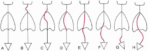
Curves are classified in three ways, based on cause, location, and direction. The causal classification for scoliosis is the most accepted method of categorizing lateral spinal column deviations. (1) This method of classification includes two major descriptions of scoliosis, identifying them as either structural or non-structural. Within these two major divisions, the specific origin of the scoliosis is then identified. (Table 4-1) The location classification is based on the region of the spine in which the apex vertebra is located and describes the curvature. (Fig. 4-1; Table 4-2) Classification based on direction describes the plane into which the concavity of the curve projects, for example, dextroscoliosis ( patient’s right), levoscoliosis ( patient’s left), lordoscoliosis ( patient’s anterior), and kyphoscoliosis (patient’s posterior). Throughout this chapter we use the causal classification of scoliosis.


Table 4-1 Causal Classification of Scoliosis


Structural scoliosis


I.


Idiopathic


A.


Infantile


1.


Resolving


2.


Progressive


B.


Juvenile (3-10 years)


C.


Adolescent (> 10 years)


II.


Neuromuscular


A.


Neuropathic


1.


Upper motor neuron


a.


Cerebral palsy


b.


Spinocerebellar degeneration


(1)


Friedreich’s disease


(2)


Charcot-Marie-Tooth disease


(3)


Roussy-Levy disease


c.


Syringomyelia


d.


Spinal cord tumor


e.


Spinal cord trauma


f.


Other


2.


Lower motor neuron


a.


Poliomyelitis


b.


Other viral myelitides


c.


Traumatic


d.


Spinal muscular atrophy


(1)


Werdnig-Hoffmann


(2)


Kugelberg-Welander


e.


Myelomeningocele (paralytic)


3.


Dysautonomia (Riley-Day)


4.


Other


B.


Myopathic


1.


Arthrogryposis


2.


Muscular dystrophy


a.


Duchenne’s (pseudo-hypertrophic)


b.


Limb-girdle


c.


Facioscapulohumeral


3.


Fiber-type disproportion


4.


Congenital hypotonia


5.


Myotonia dystrophica


6.


Other


III.


Congenital


A.


Failure of formation


1.


Wedged vertebra


2.


Hemivertebra


B.


Failure of segmentation


1.


Unilateral (unsegmented bar)


2.


Bilateral


C.


Mixed


IV.


Neurofibromatosis


V.


Mesenchymal disorders


A.


Marfan’s


B.


Ehlers-Danlos


C.


Other


VI.


Rheumatoid disease


VII.


Trauma


A.


Fracture


B.


Surgical


1.


Postlaminectomy


2.


Post-thoracoplasty


C.


Irradiation


VIII.


Extraspinal contractures


A.


Postempyema


B.


Postburn


IX.


Osteochondrodystrophies


A.


Diastrophic dwarfism


B.


Mucopolysaccharidoses (e.g., Morquio’s syndrome)


C.


Spondyloepiphyseal dysplasia


D.


Multiple epiphyseal dysplasia


E.


Other


X.


Bone infection


A.


Acute


B.


Chronic


XI.


Metabolic disorders


A.


Rickets


B.


Osteogenesis imperfecta


C.


Homocystinuria


D.


Other


XII.


Related to lumbosacral joint


A.


Spondylolysis and spondylolisthesis


B.


Congenital anomalies of the lumbosacral region


XIII.


Tumors


A.


Vertebral column


1.


Osteoid osteoma


2.


Histiocytosis X


3.


Other


B.


Spinal (see II. Neuromuscular)


Non-structural scoliosis


I.


Postural


II.


Hysterical


III.


Nerve root irritation


A.


Herniation of the nucleus pulposus


B.


Tumors


IV.


Inflammatory (e.g., appendicitis)


V.


Related to leg length discrepancy


VI.


Related to contractures about the hip


Figure 4-1 CLASSIFICATION OF SCOLIOSIS ACCORDING TO LOCATION. A, Normal spine; B, cervical; C, cervicothoracic; D, midthoracic; E, thoracolumbar; F, lumbar; G, lumbosacral; H, double lumbar and thoracic.


Table 4-2 Location Classification of Scoliosis


Classification


Apex Vertebra


Cervical


C1-C6


Cervicothoracic


C7-T1


Thoracic


T2-T11


Thoracolumbar


T12-L1


Lumbar


L2-L4


Lumbosacral


L5-S1


Structural

A structural scoliosis is a lateral curvature that is rigid and fails to correct on recumbent or lateral-bending radiographic studies. Many disorders cause a structural scoliosis, including idiopathic, congenital segmentation anomalies, neuromuscular, neurofibromatosis, and others. (Table 4-1)


Idiopathic

Idiopathic scoliosis is the most common form of lateral spinal deviation, accounting for up to 80% of scoliosis. (2) The cause is unknown, although many factors have been implicated, including connective tissue disease, diet, enzymes, muscular imbalance, vestibular dysfunction, and inheritance. (3,4) Of all possible causes, an inherited genetic defect appears to play a significant role; up to 30% of patients have another family member with significant scoliosis. (5,6,7) A positive family history does not translate into worse curves or progressive curves. (7) In idiopathic scoliosis, < 2% of the thoracic curves occur to the left, but when present they are more common in females than in males and have a higher incidence of underlying pathology. (8)

The onset for idiopathic curves distinctively occurs during growth periods, which allows for further classification based on age. This age-based classification categorizes the curves as infantile, juvenile, or adolescent idiopathic scolioses.


Infantile Idiopathic Scoliosis.

The infantile form occurs between birth and 3 years of age. The majority will disappear (resolving infantile idiopathic scoliosis), but some will progress (progressive infantile idiopathic scoliosis). (9) The progressive form is rare in the United States, is slightly more common in males, and is usually a left convex thoracic curve.


Juvenile Idiopathic Scoliosis.

The onset of juvenile idiopathic scoliosis is between 3 and 10 years of age, with an average age of 7 years. There is female gender predominance of 4 to 1. (10) Thoracic curves are the most common type. As many as 30% will eventually require corrective surgery.


Adolescent Idiopathic Scoliosis.

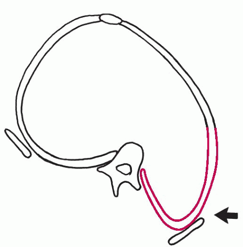
The adolescent form is the most common type of idiopathic scoliosis. Females are predominantly affected, in a ratio of 9 to 1, and the most frequent curve is the right thoracic convexity. (11) The curvature develops in the period between the age of 10 and skeletal maturity, and the curvature is often discovered during the screening of schoolchildren with the Adam’s test. If during the Adam’s test forward flexion reveals a rib hump, a structural scoliosis with rotation should be suspected. (Fig. 4-2) The critical time period for progression,
which may be rapid, is between the ages of 12 and 16 years. (Fig. 4-3) Once spinal growth has ceased, as indicated by fusion of the iliac apophysis, further rapid progression is unlikely, although slow progression of idiopathic curves throughout adulthood is a recognized complication.
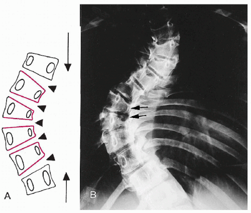


Figure 4-2 RIB HUMP OWING TO SCOLIOSIS. Note the posterior vertebral body rotation on the side of the rib hump (arrow), which is usually most prominent on the convex surface at the apex of the curve. This correlates with the physical observation of a persistent rib hump on forward flexion (Adam’s position) characteristic of a structural scoliosis.


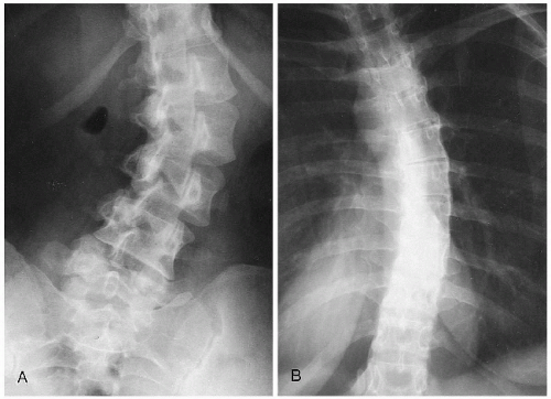
Figure 4-3 IDIOPATHIC SCOLIOSIS. A. AP Lumbar Spine. The abnormality in this 25-year-old female was first detected when she was 12 years of age. Despite bracing, her scoliosis progressed and then remained unchanged as shown here. B. AP Thoracic Spine. Observe the right convex midthoracic scoliosis in this 23-year-old female; it was clinically silent and fortuitously discovered during a chest radiographic examination. COMMENT: Many idiopathic scolioses remain asymptomatic and may be found as an incidental finding in adulthood. Cosmetic deformity may be slight despite even relatively large magnitude scoliotic curvatures.

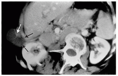
The natural history of scoliosis has not significantly changed over the years. (12) Later in adult life superimposed degenerative changes may allow the curvature to increase on an average of 15° (13) and occasionally may create nerve entrapment syndromes. (14) Generally, curves of greater magnitude at skeletal maturity show greater progression through adulthood. In severe curves, iliocostal pain may occur secondary to rib impingement onto the iliac crest. (Fig. 4-4)

Other findings in idiopathic scoliosis are the development of lateral wedge deformities of the vertebral bodies within the apex of the curve. These wedge deformities persist into adult life and are the result of excessive compressive forces that impair the growth at the discovertebral junction on the concave side of the curvature (Hueter-Volkmann principle). (Fig. 4-5) Three other curves that are frequently present are a right thoracolumbar, a left lumbar, and a combined form of left lumbar and right thoracic. (2,15) The normal psoas shadow on frontal radiographs is often not visualized on the concave side of a lumbar scoliosis. (16)

There is a 10 times greater incidence of congenital heart disease than in the general population when the idiopathic curve is > 20°. (17) The lumbar lordosis, thoracic kyphosis, and cervical lordosis are often reduced, and an increased incidence of co-existing pes cavus has been noted. (18)


Figure 4-4 ADVENTITIOUS BURSA, ILIOCOSTAL IMPINGEMENT. CT, Axial Subcostal Region. Observe the fluid density soft tissue mass between the ribs and the iliac crest, where impingement was occurring from an adventitious bursa (arrows). COMMENT: This 45-year-old female patient had suffered severe thoracolumbar scoliosis of at least 60° since adolescence and ambulated with a walking aid. She then complained of anterolateral pain near the tips of the eleventh and twelfth ribs. After the CT study a single injection of corticosteroid under ultrasound guidance relieved all pain, and a follow-up CT 4 weeks later confirmed its resolution.


Figure 4-5 HUETER-VOLKMANN PRINCIPLE. A. AP View. Excessive compressive axial loading at the discovertebral junction (arrowheads) owing to scoliosis during the period of bone growth may produce permanent wedge-shaped vertebral bodies. B. AP Thoracic Spine. The four vertebral bodies at the apices of the curves are notably wedged. Also observe the pedicle migration of these segments, signifying vertebral rotation. (Courtesy of Leo C. Wunsch Sr, DC, DACBR, Denver, Colorado.)


Congenital

Congenital scoliosis is distinguished by associated anomalies of the vertebrae or ribs. The most frequently observed anomalies are hemivertebrae, block vertebrae, spina bifida, bridging vertebral bars, joint deformities, fusion of ribs, and other rib malformations. (19) (Fig. 4-6) The scoliosis is typically a short C-shaped curve and may be rapidly progressive during growth spurts. Occasionally, anterior vertebral body defects may cause superimposed kyphosis (kyphoscoliosis). (20) There is a frequent association of genitourinary system anomalies with congenital scoliosis. (21)


Neuromuscular

A large spectrum of neuropathic and myopathic disorders may produce a progressive spinal deformity. (Table 4-1) Neuropathic scoliosis is distinctively a long C-shaped curve, frequently extending from the sacrum to the lower cervical region. The most common neuropathy associated with scoliosis is poliomyelitis. (22) The convex side is oriented toward the unaffected muscle group. Intersegmental rotation may be severe in these curves, and rapid progression in the curvature angle frequently occurs between the ages of 12 and 16 years. Cerebral palsy produces the same type of long C-shaped curve. Other neurologic disorders associated with scoliosis include syringomyelia, spinal cord tumor, trauma, and dysautonomia. A scoliosis of 15° or more occurring before the age of 11 should be viewed with a high index of suspicion for underlying intraspinal pathology. (23) (Fig. 4-7) Left-sided thoracic curves similarly have a higher incidence of intraspinal pathology, including tumors of the spinal cord or vertebrae, syringomyelia, and Arnold-Chiari malformations, all of which are best evaluated with MRI. (24,25)

Myopathic scoliosis is usually of the long C-shaped curve configuration. The most frequent cause is muscular dystrophy of Duchenne. An increasing lordosis usually precedes the onset of the scoliotic deformity. (26) The scoliosis that forms is often rapidly progressive and severe, requiring fusion and rod instrumentation. Once the patient has been confined to a wheelchair, the formation of a scoliosis is almost inevitable. (27)


Neurofibromatosis

Neurofibromatosis is an inherited congenital disorder of neuroectodermal and mesodermal origin tissues. The first description of the relationship between the formation of nerve and skin tumors was given by von Recklinghausen in 1882. (28) Scoliosis was first associated with neurofibromatosis in 1921 by Weiss. (29) The classic triad of diagnostic findings is (a) multiple, soft, elevated cutaneous tumors (fibroma molluscum); (b) cutaneous
pigmentation (café-au-lait spots); and (c) neurofibromas of peripheral nerves. In addition, various skeletal lesions, including erosions, intraosseous cystic defects, deformity, pseudo-arthrosis, growth aberrations, and cranial abnormalities, may be present in up to 50% of these patients. (30)
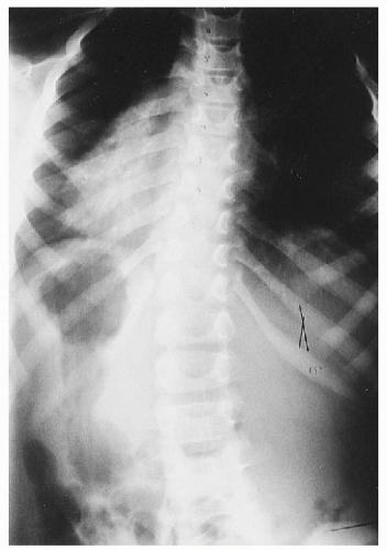


Figure 4-6 CONGENITAL SCOLIOSIS. A. Hemivertebra AP Lumbar Spine. Observe the two pedicles at the apex of this thoracolumbar scoliosis, caused by a fused hemivertebra (arrows). B. Lumbar Hemivertebra. An unfused hemivertebra that has precipitated a structural scoliosis (arrow) is demonstrated. C. Hemivertebra with Disc Degeneration, AP Lumbar Spine. Note the fused hemivertebra at the apex of this midlumbar scoliosis (arrows). Observe the severe disc degeneration of the lower lumbar segments with exuberant osteophytes, sclerosis, and loss of disc space, which is maximal on the concave side of the scoliosis. D. Synostosis, AP Ribs. Observe the thoracolumbar scoliosis as a result of a congenital localized lack of separation at the eleventh and twelfth ribs (arrow). (Panel A courtesy of Russell Banks BAppSc (Chiro), Melbourne, Australia.)


Figure 4-7 LEFT THORACIC SCOLIOSIS. AP Thoracolumbar Spine. Observe the long segment, C-shaped scoliosis convex to the left as evident by the relative positions of the heart and gastric air bubble. COMMENT: Left thoracic curves in patients under the age of 11 years may be a marker for underlying spinal canal tumors, syringomyelia, posterior fossa brain tumors, and Arnold-Chiari malformations. MRI of the entire spinal cord, including the posterior fossa, should be performed on the basis of this finding. (Courtesy of Anne P. Odenweller, DC, Baton Rouge, Louisiana.)

Scoliosis is the most common bony abnormality in neurofibromatosis patients and is present in 10-50% of cases. (30,31,32) Varying degrees of scoliosis occur, from mild to severely deforming angulations. (Figs. 4-8 and 4-9) The most conspicuous features, when present, consist of a short, angular deformity with dysplasia of the vertebral bodies. Notably, the scoliosis frequently progresses and requires surgical fusion for stabilization. Kyphosis is the most common superimposed deformity. Additional findings that suggest neurofibromatosis-induced scoliosis are enlarged foramina, posterior and lateral vertebral body scalloping, deformed ribs (twisted ribbons), and an adjacent smooth paraspinal soft tissue mass owing to either a neurofibroma or a protruding lateral meningocele.


Miscellaneous Causes


Infection.

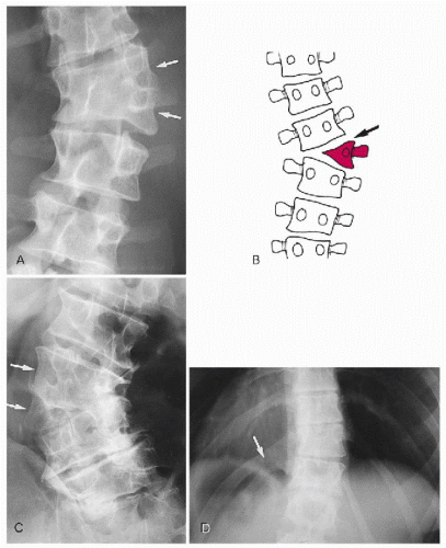
Infectious processes, such as tuberculosis, may precipitate spinal deformity as a result of collapse and bony destruction. (Fig. 4-10) The most distinctive deformity is a sharp, angular kyphosis (gibbus), although varying degrees of scoliosis may also occur.


Radiation.

Irradiation of the growing spine may produce vertebral abnormalities in up to 75% of patients. (33) These changes consist of growth arrest lines, endplate irregularities, altered vertebral shape, and scoliosis. The most commonly irradiated childhood disorders are Wilms’ tumor in the kidney and neuroblastoma in the adrenal gland. The focal point of the irradiation is the organ of involvement; however, the adjacent spine and iliac bone are frequently overlapped and affected by the treatment. This overlap produces the classic tandem findings of a small iliac wing, small vertebral bodies, and a lumbar scoliosis with a convexity away from the irradiated side. (Fig. 4-11) Two types of lumbar spine deformity can occur: a mobile scoliosis and a fixed rotary scoliosis caused by unilateral shortened laminae and pedicles. (34) In both types of curves, the convexity is away from the side of irradiation.


Trauma.

Injuries to the spine that produce fracture or dislocation may also induce a lateral spinal curvature, which may be permanent. (Fig. 4-12) Most spinal injuries produce curves that are concave to the side of injury; however, transverse process fractures are unique in that they commonly produce curves that are convex to the side of fracture. Although other spinal injuries are compressive in nature, transverse process fractures are avulsions created by the attached musculature. (35)


Spondylolisthesis.

Scoliosis found in association with lumbar spondylolisthesis is common, found in almost 25% of cases. The cause is often difficult to determine, but in 30% of
cases, the scoliosis may be the result of greater slippage of the spondylolytic vertebra on one side (olisthetic scoliosis). (36) (Fig. 4-13) A more common cause of scoliosis, seen in at least 40% of spondylolisthesis cases, is muscle spasm caused by pain. (37) Scoliosis in asymptomatic spondylolisthesis occurs in 6% of cases. (37)
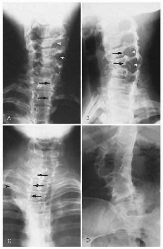


Figure 4-8 NEUROFIBROMATOSIS. A. AP Lower Cervical Spine. Observe the mild cervicothoracic scoliosis; the most dramatic changes are foraminal expansion (arrowheads) and lateral vertebral scalloping (arrows). B. Oblique Cervical Spine. Note that the foramina are greatly expanded from marked dural ectasia (arrowheads); note also the posterior scalloping of the vertebral bodies (arrows). C. AP Upper Thoracic Spine. Observe the upper thoracic curvature, lateral body scalloping (arrows), and the paraspinal mass (arrowhead) of an associated meningocele. D. AP Lumbar Spine. Observe the bizarre distortion of the lumbar bodies, posterior arches, and intervertebral foramina accompanying the scoliosis. COMMENT: Scoliosis is a common finding in neurofibromatosis and is often associated with kyphosis. (Panels A and B courtesy of William E. Litterer, DC, DACBR, Fellow, ACCR, Elizabeth, New Jersey; panel C courtesy of Clayton F. Thomsen, DC, Sydney, Australia; panel D courtesy of Lawrence A. Cooperstein, MD, Pittsburgh, Pennsylvania.)

Only gold members can continue reading. Log In or Register to continue

Stay updated, free articles. Join our Telegram channel

Aug 2, 2016 | Posted by in GENERAL RADIOLOGY | Comments Off on Scoliosis

Full access? Get Clinical Tree

Get Clinical Tree app for offline access