Abstract
Segmental and lobar consolidations are the result of alveolar filling in contrast with lobar or segmental atelectasis, which is the result of alveolar collapse. Atelectasis results in loss of lung volume while consolidation maintains or even increases lung volume. Consolidations are most often caused by pneumonias but may result from obstructive pneumonia distal to an endobronchial tumor, peripheral invasive mucinous adenocarcinoma, or lymphoma.
Keywords
aspiration pneumonia, bronchopneumonia, drown lung, infarction, invasive mucinous adenocarcinoma, lobar pneumonia, lung torsion, lymphoma, obstructive pneumonia, tuberculosis
Questions
- 1.
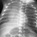
Homogeneous, left lower lobe consolidation, as seen in Fig. 14.1, A and B , is the typical radiologic manifestation of community-acquired lobar pneumonia. What is the most likely causal agent?
- a.
Mycoplasma pneumoniae.
- b.
Streptococcus pneumoniae.
- c.
Mycobacterium avium intracellulare.
- d.
Legionella pneumophilia.
- e.
Klebsiella pneumoniae.
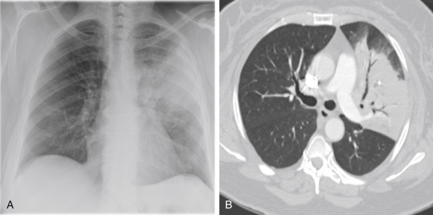
Fig. 14.1
- a.
- 2.
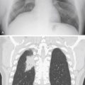
Refer to Fig. 14.2, A and B . An expanded lobe with interspersed lucent spaces (shown by the black arrows on the figure), which are suggestive of multiple cavities, is a classic appearance for pneumonias caused by which of the following agents?
- a.
S. pneumoniae.
- b.
L. pneumophilia.
- c.
K. pneumoniae.
- d.
M. pneumoniae.
- e.
Adenovirus.

Fig. 14.2
- a.
- 3.
The combination of hilar adenopathy and right middle lobe consolidation in a 15-year-old should suggest which diagnosis?
- a.
M. pneumoniae.
- b.
Gram-negative pneumonia.
- c.
S. pneumoniae.
- d.
Respiratory syncytial virus.
- e.
Primary tuberculosis.
- a.
- 4.
Which type of lung cancer is most likely to cause an expanded lobe?
- a.
Invasive adenocarcinoma.
- b.
Small cell carcinoma.
- c.
Large cell carcinoma.
- d.
Squamous cell carcinoma.
- e.
Semi-invasive adenocarcinoma.
- a.
Discussion
Lobar Consolidation
Lobar consolidation results from alveolar filling with fluid, exudate, or tumor that solidifies the lung. 221 The radiologic appearance of a consolidated lobe is a homogeneous confluent opacity that obliterates the normal vascular markings and often contains air bronchograms (see Fig. 14.1, A and B ). Lobar pneumonia is the most common cause of lobar consolidation ( Charts 14.1 and 14.2 ). The opacity is frequently bounded by the fissures, and stretched fissures may even lead to the appearance of an expanded lobe (see Fig. 14.2, A and B ). Because the airways are frequently air filled and surrounded by airless lung, air bronchograms are another component of the classic appearance of lobar consolidation.
- I.
Lobar pneumonia
- II.
Bronchopneumonia
- A.
Pseudomonas 50
- B.
K. pneumoniae
- C.
Bacillus proteus
- D.
Escherichia coli
- E.
Anaerobes ( Bacteroides and Clostridia )
- F.
Legionella pneumophila 140 , 461 , 498
- G.
Staphylococcus aureus
- H.
Nocardiosis 50 , 208 and actinomycosis 164 , 300
- I.
S. pneumoniae 286
- J.
Serratia 28
- A.
- III.
Aspiration pneumonia
- IV.
Tuberculosis 388 , 528 and atypical mycobacteria
- V.
Pulmonary embolism 266
- A.
Hemorrhage and edema
- B.
Infarction
- A.
- VI.
Neoplasms
- A.
Obstructive pneumonia (endobronchial tumor)
- B.
Invasive mucinous adenocarcinoma (formerly bronchioloalveolar cell carcinoma) 134
- C.
- A.
- VII.
Mitral regurgitation with pulmonary edema localized to the right upper lobe 213
- VIII.
- I.
Streptococcus pneumoniae 175
- II.
Klebsiella pneumoniae 50
- III.
Pseudomonas 50
- IV.
Staphylococcus 50
- V.
Tuberculosis 528
- VI.
Carcinoma with obstructive pneumonia (drowned lung)
From the foregoing description, the distinction of lobar consolidation from atelectasis would appear to be simple. Fraser et al. 175 have emphasized that consolidation of a lobe with fluid is the antithesis of lobar atelectasis. Therefore, bulging fissures that indicate lobar expansion are unequivocal evidence of consolidation. As mentioned in Chapter 13 , the displacement of fissures together indicates volume loss in the involved lobe. The problem with applying this approach to the evaluation of a lobar opacity for the purpose of distinguishing atelectasis from consolidation is not in the evaluation of the lobe that is expanded, but rather in the evaluation of the lobe of diminished size. It will become apparent in this discussion that a number of the processes that consolidate the lung also produce small airway obstruction, so that the volume loss produced by the process may be greater than the quantity of fluid that is consolidating the lung. Thus, a consolidating process may also be the cause of atelectasis. For example, bronchopneumonia, which causes air space consolidations, also leads to small airway obstructions that may result in atelectasis.
Lobar Pneumonia
The distinction between lobar pneumonia and bronchopneumonia has been deemphasized in favor of a bacteriologic classification of pneumonias, which is more relevant in determining appropriate therapy. However, knowledge of the gross morphologic distinctions between the two types of pneumonia is important for understanding the variety of radiologic patterns that may be encountered in acute pneumonias.
The pathogenesis of lobar pneumonia is the key to understanding the spectrum of radiologic patterns. Experimental evidence indicates that the pathogens, in the form of small, infected, mucous particles, are inhaled to the periphery of the lung, where they set up foci of reaction. Initially, the tissue reaction involves the exudation of watery edema fluid into alveolar spaces. The exudate then spreads into the air passages and their alveoli. As the alveoli fill, the exudate spreads into adjacent lobules and segments. This movement of the exudate occurs via the pores of Kohn, canals of Lambert, and small airways, but does not appear to spread via the bronchovascular bundle or interstitium of the lung. The watery edema fluid serves as a culture medium for the rapid multiplication of the bacteria. The alveolar walls respond to the organisms by releasing polymorphonuclear leukocytes. The spread of the process through the collateral channels, rather than the bronchioles, explains why lobar pneumonia often does not follow a segmental distribution. Rather, lobar pneumonia produces opacities that appear to involve multiple segments early in the course of the process. During these early phases, the radiologic appearance is that of a nonsegmental sublobar consolidation, which may appear rather sharply circumscribed because of the uniform involvement of contiguous alveoli. This leads to so-called round pneumonia. 608 Round pneumonia is more commonly seen in children but may occur as an early stage of lobar pneumonia in adults ( Fig. 14.3, A and B ). Fully developed, classic lobar consolidation is less commonly encountered because early diagnosis of bacterial pneumonia followed by appropriate antibiotic therapy frequently arrests the process in its early phases. Since the initial focus of infection is the periphery of the lung, it is not surprising that pleural effusion and empyema are potential complications. However, the occurrence of these complications is also drastically reduced by early antibiotic therapy.