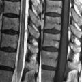
6 Sellar/Parasellar Neoplasms Pituitary adenomas are a frequent source of incidental findings, warranting serial MRI examinations. Three-fourths, however, are hormonally active and thus brought to early clinical attention. Microadenomas (<10 mm in diameter) appear as low to moderate SI focal lesions on T1WI, with variable SI on T2WI (Fig. 6.1A). This appearance, visualized against the moderate SI of the pituitary, render microadenomas often difficult to visualize without contrast. Early (<5 minutes) postcontrast MRI well demonstrates the minimally enhancing adenoma against the brightly enhancing pituitary gland (Figs. 6.1B,C, white arrows). On delayed postcontrast scans, the adenoma may be iso- to hyperintense to the gland. Contrast administration is essential preoperatively and when Cushing disease is suspected, as ACTH (adrenocorticotrophic hormone) secreting tumors tend to be the smallest of the microadenomas. MRI is not useful for distinguishing the various types of adenomas, although both prolactinomas—the most common functioning adenoma— and growth hormone-secreting adenomas tend to occur in the lateral aspects of the gland. 3 T MRI offers substantial advantages for imaging of pituitary microadenomas, making possible acquisition of images with a slice thickness of 2 mm.
Stay updated, free articles. Join our Telegram channel

Full access? Get Clinical Tree


