Imaging
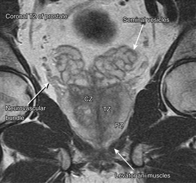
Traditionally, the seminal vesicles were evaluated with seminal vesiculography. This has largely been replaced by computed tomography (CT), magnetic resonance imaging (MRI), and ultrasound ( Figure 74-1 ).

Computed Tomography
The seminal vesicles are of soft tissue attenuation ( Figure 74-2 ). Cysts and small masses that do not deform the seminal vesicle are not well seen. Large masses or inflammatory change associated with infection or abscess can be appreciated. Calcification is clearly seen.

Magnetic Resonance Imaging
MRI is a valuable tool in evaluating the seminal vesicles owing to its multiplanar imaging capabilities and superb soft tissue contrast resolution ( Figure 74-3 ). It clearly demonstrates cystic lesions and is more accurate for staging of solid neoplasms than CT or ultrasound.

Ultrasound
Transrectal ultrasound (TRUS) is rapid, inexpensive, and generally well tolerated. It is superior to CT because it better delineates the internal structure of the seminal vesicles. It also does not require the administration of ionizing radiation and can be used to guide seminal vesicle biopsy or aspiration.
Specific Lesions
Seminal Vesicle Cyst
Etiology
Seminal vesicle cysts may be congenital or acquired. They manifest as a mass between the rectum and base of the bladder and may be associated with ejaculatory duct dilation. They are usually unilateral, unilocular, and resemble dilated seminal vesicles ( Figure 74-4 ). Congenital cysts are often associated with ipsilateral genitourinary anomalies. An acquired cyst is usually associated with ejaculatory duct or seminal vesicle inflammation or obstruction such as prostatitis, seminal vesiculitis, or prostate surgery.

Associations with seminal vesicle cysts include the following:
- •
Autosomal dominant polycystic kidney disease in which the seminal vesicle cysts are usually bilateral
- •
Invasive local tumor or primary tumor of the seminal vesicle
- •
Infection or chronic prostatitis
- •
Benign prostatic hypertrophy
- •
Ejaculatory duct obstruction
Clinical Presentation
Seminal vesicle cysts usually are asymptomatic but may manifest as obstruction, recurrent urinary tract infections (including epididymitis), and hematospermia.
Pathology
Seminal vesicle cysts can occur in any part of the gland. They vary in size and are benign. Superimposed infection may be seen.
Imaging
Computed Tomography.
Cysts are well-defined hypoattenuating lesions on CT.
Magnetic Resonance Imaging.
The MRI features of seminal vesicle cysts are similar to those of cysts in other locations, with low signal intensity on T1-weighted images and a unilocular smooth-walled lesion with uniform high intensity and a well-defined margin on T2-weighted images (see Figure 74-4 ). Hemorrhagic cysts have high signal intensity on both T1- and T2-weighted images.
Ultrasound.
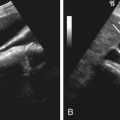
Cysts are seen as anechoic masses ( Figure 74-5 ). TRUS can be used to guide needle placement for drainage or contrast studies to more fully delineate the lesion.

Differential Diagnosis
The main differential diagnosis is müllerian duct cyst.
Treatment
No medical treatment is required for seminal vesicle cysts. When seminal vesicle cysts are large, they may cause pain and local obstruction. Draining such cysts may relieve discomfort.
Seminal Vesicle Obstruction
Etiology
Seminal vesicle obstruction is defined as a seminal vesicle with an anteroposterior diameter of more than 15 mm, length longer than 50 mm, and large anechoic areas containing sperm on aspiration. Seminal vesicle obstruction may be congenital because of an ectopic ureter or acquired secondary to a local mass. It is important to recognize because it is associated with pain and male infertility.
Clinical Presentation
The obstruction may be asymptomatic or painful when the gland becomes enlarged.
Pathology
When seminal vesicle obstruction is due to congenital causes, it is most often unilateral. When acquired, it may be unilateral or bilateral. The seminal vesicles are usually normal histologically but may become secondarily infected.
Imaging
Computed Tomography.
CT will show an enlarged seminal vesicle.
Magnetic Resonance Imaging.
The enlarged seminal vesicle is of normal signal intensity unless infected or infiltrated by tumor.
Ultrasound.
Ultrasound will show an enlarged seminal vesicle.
Differential Diagnosis
No clinical or laboratory data aid in the diagnosis. Tumor marker levels may be elevated when the obstruction is caused by a malignant mass.
Treatment
There is no medical treatment for seminal vesicle obstruction. Surgery or image-guided aspiration may be required to treat symptomatic obstruction.
Seminal Vesiculitis and Abscess
Etiology
Infection of the seminal vesicles is most often secondary to acute or chronic prostatitis or acute epididymitis, although it may occur independently. The anaerobic organisms that cause prostatitis are the usual cause of seminal vesiculitis. Mycobacterium tuberculosis and Schistosoma are the most common causes in developing countries. Seminal vesiculitis may progress to a seminal vesicle abscess. Like prostate abscess, seminal vesicle abscess is more frequent in those with diabetes mellitus, chronic indwelling urinary catheter, and recent urinary tract intervention. Seminal vesiculitis and abscess may be associated with cysts and calcification of the seminal vesicles. Chronic bacterial vesiculitis is rare and difficult to diagnose.
Clinical Presentation
The cause of seminal vesiculitis is similar to other causes of pelvic infection such as prostatitis or cystitis; these infections may occur simultaneously.
Pathology
The disorder is most often unilateral but may be bilateral. Infection and inflammation are found on histologic examination.
Imaging
Computed Tomography.
In the acute setting, the seminal vesicles may appear normal or asymmetrically enlarged. Surrounding inflammation also may be visualized. When abscess formation occurs, the involved seminal vesicle is enlarged, of low attenuation, or fluid-filled. Chronic infection causes wall thickening, contraction, and intraluminal or wall calcification on cross-sectional images.
Magnetic Resonance Imaging.
Seminal vesiculitis and seminal vesicle abscess demonstrate low signal intensity on T1-weighted images. Signal intensity is higher than the normal seminal vesicle on T2-weighted images. Both enhance with gadolinium.
Ultrasound.
In vesiculitis, TRUS may demonstrate enlargement of seminal vesicles (>14 mm thick) and thickened septa. Seminal vesicle abscess has features similar to those of abscesses elsewhere—a collection of heterogeneous but predominantly low echogenicity.
Imaging Algorithm.
Seminal vesiculitis usually does not require investigation unless treatment fails, infection recurs, or there is concern that a mass is present.
Differential Diagnosis
Seminal vesicle abscesses and infection are diagnosed clinically by a positive ejaculate culture. Imaging is not usually required in the investigation of infection.
Treatment
Antibiotic therapy is the first-line treatment for seminal vesiculitis. A short trial of antibiotic treatment may be performed for small abscesses. If the seminal vesicle abscess fails to respond rapidly to antibiotics, transrectal or perineal aspiration and, if necessary, surgery to remove the seminal vesicles may be required.
Malignant Tumors of the Seminal Vesicles
Etiology
Primary malignant tumors are rare and usually unilateral. Adenocarcinoma and sarcoma have been reported. Secondary malignancy of the seminal vesicles is much more common and may be due to hematogeneous spread or local invasion by malignant tumors of the prostate, rectum, or bladder ( Figures 74-6 and 74-7 ). Locally invasive tumor is more likely to involve both seminal vesicles, and it may be difficult to identify the organ of origin.


Prevalence and Epidemiology
Primary seminal vesicle adenocarcinoma is very rare. It manifests in men older than 50 years of age. Sarcoma of the seminal vesicles is even less common.
Clinical Presentation
The diagnosis of primary seminal vesicle neoplasm can be difficult because these lesions usually do not cause symptoms until late in their course and because symptoms are often nonspecific. Presenting signs and symptoms include urinary retention, dysuria, hematuria, and hematospermia.
Pathology
Adenocarcinoma is the usual histologic type of primary seminal vesicle malignancy; however, epithelial stromal tumors, sarcomas (leiomyosarcoma, angiosarcoma, müllerian adenosarcoma-like tumor), phylloides tumor, and choriocarcinoma of the seminal vesicles are all reported. Invasion from the prostate, rectum, or other organ should be ruled out (see Figures 74-7 to 74-9 ).


Imaging
Computed Tomography.
Primary tumors are centered on and predominantly confined to the seminal vesicles, whereas secondary tumors are centered on the prostate, bladder, or rectum. However, CT cannot reliably distinguish among tumor, chronic inflammation, and amyloid.
Magnetic Resonance Imaging.
On MRI, a seminal vesicle tumor appears as a heterogeneous mass of intermediate signal on T1-weighted imaging and as heterogeneous high signal intensity on T2-weighted imaging. As with CT and TRUS, MRI cannot distinguish between benign and malignant solid masses.
Ultrasound.
Solid tumors of the seminal vesicle may be isoechoic to the prostate but relatively hyperechoic to the normal seminal vesicle. There are no imaging characteristics indicative of a solid mass being benign or malignant.
Differential Diagnosis
Because prostate cancer involving the seminal vesicle often cannot be distinguished from a primary seminal vesicle cancer, the prostate-specific antigen (PSA) level should be measured. In primary seminal vesicle malignancy, serum levels of markers for prostate cancer (i.e., PSA, prostatic acid phosphatase) are normal and the level of serum carcinoembryonic antigen is elevated. Positive staining for Ca-125 has been reported and may be useful to distinguish this from other tumors, such as those arising from the prostate or bladder.
Treatment
Advanced cancers, not amenable to surgical treatment, may be treated palliatively with chemotherapy. Seminal vesicle neoplasms manifest late and usually are not amenable to curative resection at the time of diagnosis.
Benign Tumors of the Seminal Vesicles
Etiology
Causes of benign tumors include cystadenoma, fibroma, and leiomyoma. Cystadenomas usually are an incidental finding in elderly men. They are usually multilocular and may mimic seminal vesicle cysts.
Clinical Presentation
Benign seminal vesicles are usually asymptomatic unless they cause local obstruction.
Pathology
Benign tumors are usually unilateral. The pathologic findings vary according to the causative lesion.
Imaging
Computed Tomography.
CT poorly defines the internal structure of the seminal vesicle. CT may demonstrate a unilateral or bilateral mass. Local invasion is not a feature of these benign lesions.
Magnetic Resonance Imaging.
Cystadenomas are multilocular and may mimic seminal vesicle cysts on MRI.
Ultrasound.
Cystadenomas are multilocular and may mimic seminal vesicle cysts on TRUS. Solid tumors may be hypoechoic or hyperechoic.
Differential Diagnosis
The differential diagnosis is seminal vesicle cysts.
Treatment
Masses that cannot be differentiated from primary malignant masses should be resected.
Seminal Vesicle Calcification
Etiology
Seminal vesicle calculi and calcification most commonly occur secondary to infection with Neisseria gonorrhoeae, M. tuberculosis, or Schistosoma or to seminal vesicle obstruction. Calcification is also seen in patients with diabetes.
Clinical Presentation
May be asymptomatic and incidental or manifest as pain secondary to obstruction, infection, hematospermia, or infertility.
Pathology
Seminal vesicle calcification can be classified into calcification of the seminal vesicle wall or seminal vesicle calculi. Calcification may be secondary to or complicated by infection or obstruction.
Imaging
Computed Tomography.
CT accurately demonstrates the location of seminal vesicle calcification or calculi.
Magnetic Resonance Imaging.
MRI has limited value in assessing calcifications.
Ultrasound.
TRUS may demonstrate the location and degree of calcification, owing to its echogenicity and the presence of posterior acoustic shadowing. However, shadowing from calcification in the prostate may limit the use of ultrasound.
Differential Diagnosis
The differential diagnosis for calcification are diabetes and infection.
Treatment
No medical treatment is necessary except antibiotics when concomitant infection is present. Surgical resection of a calculus may be required if it is suspected to be causing recurrent infections, obstruction, and/or infertility.
Seminal Vesicle Amyloid
Etiology
The deposition of amyloid in the seminal vesicles appears to be a phenomenon related most frequently to aging. It is common and usually a localized finding. Systemic amyloidosis is not present in most patients. It is important not to confuse seminal vesicle amyloid with a mass, metastasis, or local invasion from an adjacent prostate or rectal tumor.
Prevalence and Epidemiology
The incidence of seminal vesicle amyloid increases with age, reaching 21% in men age 75 years and older.
Clinical Presentation
Isolated seminal vesicle amyloid is usually asymptomatic, unlike systemic amyloidosis, which involves many organs and systems in the body. It has been rarely associated with hematospermia and symptomatic enlargement of the seminal vesicle.
Pathology
Seminal vesicle amyloid may be unilateral or bilateral. Local senile amyloid deposits are a common finding at autopsy. Deposits are bilateral and symmetric and occur subepithelially in the lamina propria of the seminal vesicles in aggregates varying in size from microscopic to grossly visible seminal vesicle wall thickening. When the amyloid deposits are due to systemic amyloidosis, the amyloid is located in the walls of blood vessels or within muscle rather than in a subepithelial location.
Imaging
Computed Tomography.
CT will demonstrate focal or bilateral enlargement of the seminal vesicles.
Magnetic Resonance Imaging.
Seminal vesicle amyloidosis is of low signal intensity on T2-weighted imaging, as is tumor invasion (see Figure 74-9 ). MRI may distinguish tumor invasion, although not with complete accuracy. Unlike tumor, seminal vesicle amyloid does not enhance.
Ultrasound.
Amyloid is demonstrated as areas of low echogenicity that are indistinguishable from other seminal vesicle masses. TRUS may be helpful in distinguishing adjacent organ tumor spread from a focal seminal vesicle mass.
Differential Diagnosis
A normal PSA helps rule out prostate tumor invasion. However, even if the PSA value is elevated, the seminal vesicle pathologic process may still be amyloid and not metastatic prostate cancer. Biopsy helps confirm the diagnosis and rule out local invasion from tumors of adjacent organs.
Treatment
No medical or surgical treatment is required. Because the radiologic appearance can be indistinguishable from that of tumor, TRUS-guided biopsy of the seminal vesicles should be considered in all cases of seminal vesicle mass or abnormality, particularly if the diagnosis has been made incidentally at MRI.


