Sesamoiditis
Anatomic Considerations
The sesamoid bones are small, rounded structures that are embedded in the flexor tendons of the foot and usually are in close proximity to the joints. Sesamoid bones of the first metatarsal occur in almost all patients, with sesamoid bones being present in the flexor tendons of the second and fifth metatarsals in a significant number of patients (Figs. 24.1 and 24.2). These sesamoid bones serve to decrease friction and pressure of the flexor tendon as it passes in proximity to a joint.
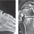
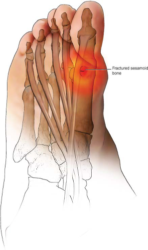 FIGURE 24.1 Sesamoiditis most commonly affects the first sesamoid bone of the first metatarsal head, although the sesamoid bones of the second and fifth metatarsal heads also are subject to the development of sesamoiditis. |
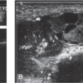
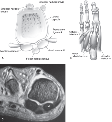 FIGURE 24.2 Illustrations of the sesamoid relationships in the axial (A) and plantar planes (B). C: Axial fat-suppressed fast SE image demonstrating the medial (M) and lateral (L) sesamoids seated in the metatarsal grooves. (From Berquist TH. Imaging of the Foot and Ankle. 3rd ed. Philadelphia, PA: Lippincott Williams & Wilkins; 2011:26.) |
Clinical Correlates
Sesamoiditis is one of the most common pain syndromes of the forefoot. Caused by inflammation of the sesamoid bones, sesamoiditis is characterized by tenderness and pain over the metatarsal heads. The first sesamoid bone of the first metatarsal head is most commonly affected, although the sesamoid bones of the second and fifth metatarsal heads also are subject to the development of sesamoiditis (Fig. 24.3). The patient suffering from sesamoiditis frequently complains that it feels like he or she is walking with a stone in his or her shoe. The pain of sesamoiditis worsens with prolonged standing or walking for long distances and is exacerbated by improperly fitting or padded shoes. Sesamoiditis is most often associated with pushing-off injuries during football or repetitive microtrauma from running or dancing.
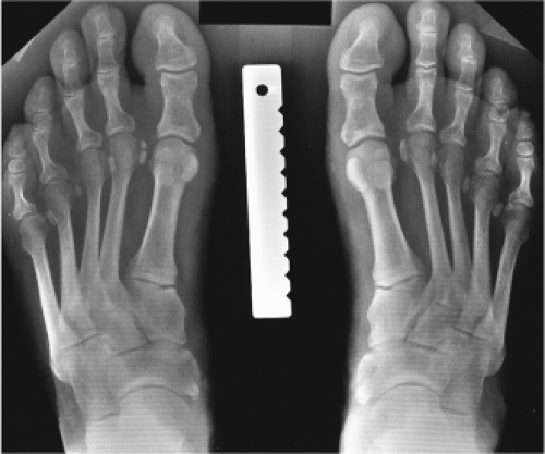 FIGURE 24.3 Standing posteroanterior views of the feet demonstrating sesamoids at all metatarsophalangeal joints. (From Berquist TH. Imaging of the Foot and Ankle. 3rd ed. Philadelphia, PA: Lippincott Williams & Wilkins; 2011:26.) |
On physical examination, the pain of sesamoiditis can be reproduced by pressure on the affected sesamoid bone. Metatarsalgia is frequently confused with sesamoiditis. In contradistinction
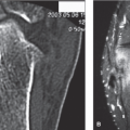
to metatarsalgia where the tender area of palpation remains over the metatarsal heads, with sesamoiditis, the tender area moves with the flexor tendon when the patient is asked to actively flex his or her toe. The patient with sesamoiditis often exhibits an antalgic gait in an effort to reduce weight bearing during walking. With acute trauma to the sesamoid, ecchymosis over the plantar surface of the foot may be present and on occasion fracture of the sesamoid bone may occur (Figs. 24.4 and 24.5).
to metatarsalgia where the tender area of palpation remains over the metatarsal heads, with sesamoiditis, the tender area moves with the flexor tendon when the patient is asked to actively flex his or her toe. The patient with sesamoiditis often exhibits an antalgic gait in an effort to reduce weight bearing during walking. With acute trauma to the sesamoid, ecchymosis over the plantar surface of the foot may be present and on occasion fracture of the sesamoid bone may occur (Figs. 24.4 and 24.5).
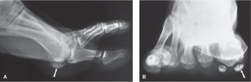 FIGURE 24.4 Lateral (A) and sesamoid (B) views demonstrating sclerosis and fragmentation of the medial sesamoid (arrow) due to fracture with avascular necrosis. (From Berquist TH. Imaging of the Foot and Ankle. 3rd ed. Philadelphia, PA: Lippincott Williams & Wilkins; 2011:194.) |
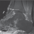
Plain radiographs are indicated in all patients who present with sesamoiditis (Fig. 24.6). Based on the patient’s clinical presentation, additional testing may be indicated, including complete blood cell count, sedimentation rate, and antinuclear antibody testing. MRI or ultrasound of the intermetatarsal bursa is indicated to help confirm the diagnosis if fracture, effusion, tendinopathy, crystal arthropathy, joint mice, synovitis, foreign body, bursitis, or ligamentous injury is suspected (Figs. 24.7 and 24.8).
Related posts:

Stay updated, free articles. Join our Telegram channel

Full access? Get Clinical Tree


