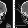
Innominate line (radiology term, tangential view of greater wing of sphenoid bone)
Hypophyseal fossa (bottom)
Nasal septum
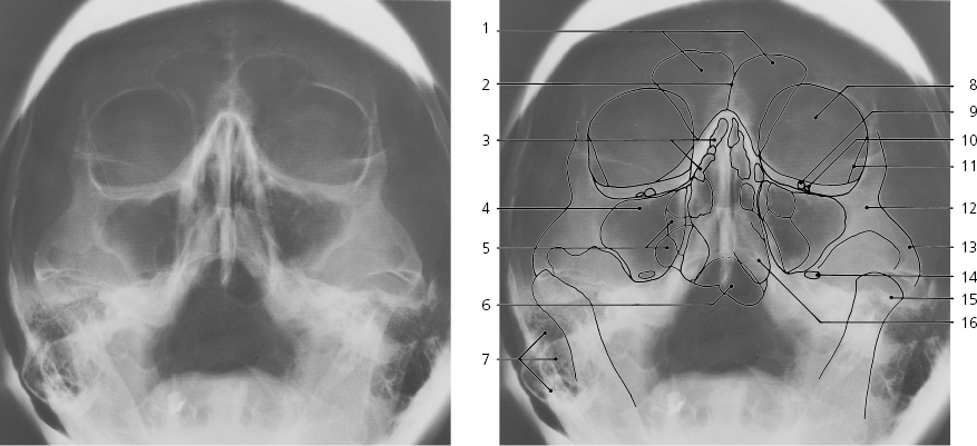
Paranasal sinuses, a-p, tilted X-ray
Frontal sinus
Septum of frontal sinus
Anterior ethmoidal air cells
Maxillary sinus
Posterior ethmoidal air cells
Sphenoid sinus
Mastoid air cells
Orbita
Foramen rotundum
Infra-orbital foramen
Innominate line (radiology term)
Body of zygomatic bone
Zygomatic arch
Oval foramen
Head of mandible
Inferior nasal concha
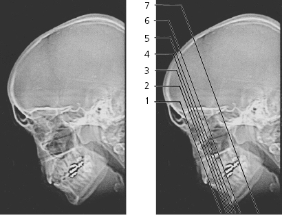
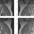
Paranasal sinuses, scout view
Lines #1–7 indicate positions of sections in the following CT series. Sections are 1 mm thick. Prone position with hyperextended neck. Sections #2–6 display the “ostiomeatal complex/unit” comprising the maxillary sinus ostium, infundibulum, uncinate process, hiatus semilunaris, ethmoidal bulla, middle concha and middle meatus. Arrows ←, → and ↔ indicate that a structure can be seen on a previous or following section or both.
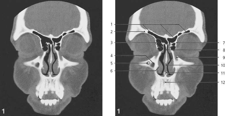
Paranasal sinuses, coronal CT
Only gold members can continue reading. Log In or Register to continue