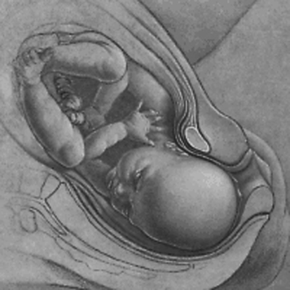
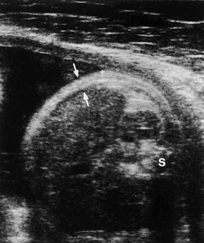
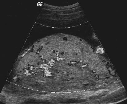
Size Greater than Dates


Size Greater than Dates


Terry J. DuBose


Obstetric sonographic examinations are frequently requested because the patient’s size is greater than the size expected for gestational age (size greater than dates) if the LMP is unknown or incorrect. The size expected for gestational age is based on the patient’s LMP and the physical examination that may include a fundal height measurement. The fundal height measurement is performed with external palpation of the uterus and measurement of the distance from the symphysis pubis to the uterine fundus. The fundal height roughly correlates in centimeters with the gestational age in weeks. This measurement is not highly accurate and may not always be a reflection of excessive fetal growth and can be affected by multiple factors, including the technique of the clinician, maternal weight, fetal position, increase in amniotic fluid, and size of the placenta. In addition, size greater than dates may be suspected when the patient has had a significant weight gain. The uterus may also present large for dates when leiomyomas are present or when ovarian masses mimic an enlarged uterus or hamper the ability to measure the uterus accurately. This chapter explores the possible results and outcomes when a patient is seen for an obstetric sonogram for size greater than dates.


Excessive Fetal Growth


Excessive fetal growth is typically divided into two categories with fetal weight in the determination. Large for gestational age (LGA) is a term suggested when the estimated fetal weight is greater than the 90th percentile for gestational age. Chapter 20 discusses the term small for gestational age, which is suggested when the estimated fetal weight is less than the 10th percentile for gestational age. Macrosomia is determined when the estimated fetal weight is greater than or equal to 4500 g. Appropriate fetal growth is clinically significant and a direct indicator of fetal well-being. Identification of fetuses that are not growing appropriately is important because as fetal growth discrepancy becomes progressively greater, whether from macrosomia or growth restriction, the risk of perinatal morbidity and mortality is significantly increased.


Estimation of Fetal Weight


The estimation of fetal weight is simply that—an estimation. Multiple variables contribute to fetal weight discrepancies. The very nature of the process for determination of fetal weight involves much variability in that sonographic measurements of the fetal head, abdomen, and femur bones, regardless of the degree of care and accuracy by the sonographer, are assigned a weight approximation on the basis of previous research data. Even the best sonographic determination of fetal weight has been estimated to be 10% discrepant of the actual weight. This potential discrepancy percentage would not appreciably affect a fetus of average size with regard to the determination of fetal weight. However, in cases of LGA, it can result in a significant discrepancy of several hundred grams. One study showed that for a confidence level of 90% that a newborn would actually weigh more than 4000 g, one must estimate the sonographic fetal weight at 4750 g.


The most reliable formula used to determine the estimated fetal weight incorporates several fetal parameters, including biparietal diameter, HC, AC, and femur length. The accuracy of this determination is associated with limitations and ranges of discrepancy. Patients and their families need to be properly educated with regard to the degree of accuracy in the estimation of fetal weight with sonographic examination.


Pregnancy Dating


Crucial information used in the proper dating of a pregnancy includes an accurate LMP date and the performance of an early baseline sonogram for later comparison. However, in about 20% to 40% of all pregnancies, the correct menstrual age is uncertain because of unknown or unclear LMP dates. Accurate assessment of the fundal height of the uterus during physical examination in cases of maternal obesity may be extremely difficult. In addition, pregnancy dating and the determination of estimated fetal weight involve the following basic assumptions:


These assumptions are averages calculated over time and across multiple populations. Just one of these assumptions may have a significant impact on the calculation of pregnancy dating. Taken together, a much wider potential range of error exists. A common source of LMP errors is due to implantation bleeding or spotting, which is mistaken for the LMP. This implantation bleeding often occurs 2 to 3 weeks after the normal LMP and leads to an estimated gestational age that is less than the true age, resulting in sonographic estimates that appear larger than dates. For these reasons, an early baseline sonographic examination is the most reliable indicator of fetal age that may be referenced throughout the duration of the pregnancy.


Macrosomia


The term macrosomia is defined as an abnormally large size of the body. The term refers to the entire fetus, neonate, or newborn. Fetal macrosomia complicates more than 10% of all pregnancies in the United States. With respect to delivery, any fetus that is too large for the maternal pelvis through which it must pass is macrosomic. The most straightforward approach to the sonographic determination of macrosomia is to use the estimated fetal weight.


Risk Factors


The major risk factor for macrosomia is gestational diabetes, which accounts for 40% of all cases. The prevalence rate of macrosomia is 25% to 42% among diabetic mothers versus 8% to 10% among nondiabetic mothers. Despite the higher frequency in diabetic mothers, nondiabetic mothers account for 60% of macrosomic cases because of their majority compared with the smaller number of diabetic mothers. Macrosomia is associated with enlargement of the placenta (Fig. 19-1; see Color Plate 17). A placental thickness obtained at a right angle to its long axis that measures greater than 3 cm before 20 weeks’ gestation or greater than 5 cm before 40 weeks’ gestation is considered abnormal.


Time of delivery is an important factor to consider. One study showed an increased incidence of macrosomia from 1.7% at 36 weeks’ gestation to 21% at 42 weeks’ gestation. Chronic and progressing macrosomia is in direct proportion to an elevated risk of associated conditions, and women who have previously delivered a macrosomic infant are at an increased risk in future pregnancies. Primary perinatal complications include shoulder dystocia, soft tissue trauma, humeral and clavicular fractures, brachial plexus injury, facial palsies, meconium aspiration, prolonged labor, and asphyxial injuries. Shoulder dystocia (Fig. 19-2) occurs when the arm of the fetus prevents or complicates delivery and may result in serious traumatic injury. Because many of these injuries are unpredictable events, available evidence suggests that planned interventions on the basis of estimates of fetal weight may not significantly reduce the incidence of shoulder dystocia and the adverse outcomes attributable to fetal macrosomia. However, because evidence strongly suggests an increased risk of prenatal complications for pregnancies with fetuses weighing greater than 4500 g, the option for cesarean delivery should be considered with fetal macrosomia in diabetic patients with small pelvic structures and in any pregnancy where macrosomia is a concern.


The sonographic fetal AC measurement has been determined to be helpful in identification of potential macrosomic infants. One study reported a less than 1% incidence rate of infant birth weights greater than 4500 g with AC measured less than 35 cm. The incidence rate significantly increased to 37% in cases where the AC was greater or equal to 38 cm. Macrosomic infants of diabetic mothers usually have organomegaly, especially a disproportional enlargement of the heart, liver, adrenals, and adipose tissue.


Hydrops fetalis is also associated with macrosomia and may manifest sonographically with one or more of the following: increased placental thickness, increased thickness of scalp or body wall greater than 5 mm (Figs. 19-3 and 19-4), hepatosplenomegaly, pleural and pericardial effusions, ascites, and structural fetal anomalies.


Stay updated, free articles. Join our Telegram channel

Aug 27, 2016 | Posted by in ULTRASONOGRAPHY | Comments Off on Size Greater than Dates

Full access? Get Clinical Tree

Get Clinical Tree app for offline access