72 Slice Excitation Order (in Fast Spin Echo Imaging)
In theory, an ideal radiofrequency pulse (RF) for MRI is uniform across the slice thickness, with sharp, distinct edges and no excitation beyond the boundaries (edges) of the slice. In practice, however, the spatial excitation of spins is invariably a distribution ranging from the nominal RF excitation flip angle at the center of the slice to largely reduced flip angles at the ill-defined edges (see Case 71). Stating this slightly differently, the flip angle varies across the slice, from one edge to the other. It also follows that there is partial excitation of tissue adjacent to, but beyond, the theoretical boundaries of the slice. Time-limited RF pulses are inherently imperfect, resulting in this nonrectangular slice profile. In multislice fast spin echo imaging, this limitation is accentuated because the slice profile is defined by the overlapping RF profiles of the excitation pulses as well as the 180° refocusing pulses applied over the duration of the echo train.
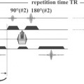
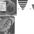
The top half of Fig. 72.1 presents the pulse diagram for a typical fast spin echo sequence. Illustrated in the bottom half of the figure, every slice receives in this instance a 90° excitation pulse, followed by multiple 180° refocusing pulses (each with a different phase encoding, Fig. 72.1, 1
Related posts:

Stay updated, free articles. Join our Telegram channel

Full access? Get Clinical Tree


