and Vincent L. Sorrell2
(1)
Division of Nuclear Medicine and Molecular Imaging Department of Radiology, University of Kentucky, Lexington, Kentucky, USA
(2)
Division of Cardiovascular Medicine Department of Internal Medicine Gill Heart Institute, University of Kentucky, Lexington, Kentucky, USA
Electronic supplementary material
The online version of this chapter (doi: 10.1007/978-3-319-25436-4_23) contains supplementary material, which is available to authorized users.
There are many conditions that impact the appearance of the small intestine and large intestine on SPECT MPI (Table23.1). The small intestine is always visualized due to clearance through the hepatobiliary system (Chamarthy and Travin2010; Gedik et al.2007). Normal peristalsis may be apparent during the course of imaging (Fig.23.1). Duodenogastric bile reflux (Fig.23.2) and intense jejunal activity may create reconstruction processing artifacts. The jejunum may herniate through the diaphragm and appear in the chest (Gedik et al.2007; Hendel et al.1999). Waiting and re-imaging usually allows for peristalsis to clear the problematic jejunal activity. An unusual case of focal “hot” activity due to a duodenal leiomyosarcoma has been reported; this case used201Tl chloride, but the99mTc radiopharmaceuticals could show this pattern after the physiologic intraluminal activity clears (Shuke et al.1995).


Table 23.1
Differential diagnosis of “hot” and “cold” imaging findings related to the small intestine and large intestine
Organ system | “Hot” finding | “Cold” finding | References |
|---|---|---|---|
Small intestine and large intestine | Herniation Stasis/slow peristalsis Neoplasm, primary (e.g., duodenal leiomyosarcoma) Previous radiopharmaceutical | Barium | Chamarthy and Travin (2010) Gedik et al. (2007) Hendel et al. (1999) Shuke et al. (1995) |
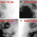
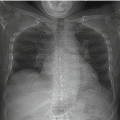
Fig 23.1


Normal small bowel peristalsis. A obese 39-year-old female underwent a normal pharmacologic stress test. As expected on a 1-day rest/stress protocol, there is more small intestinal activity on the stress images compared to the rest images (a–d). The normal gallbladder and the normal left kidney are visualized (a–d). There is duodenogastric reflux and retained liver activity, more evident on stress images, requiring tight limits for the reconstruction (e). A small, partially reversible inferolateral-apical perfusion defect is likely related to a “hot” stomach and/or a “hot” liver processing artifact (e). On gated SPECT images, the liver and stomach activity is much more apparent (f). There is preserved wall motion and wall thickening, and normal global left ventricular function with a LVEF of 57 %. Note that the large pendulous “cold” breasts seen on the raw data (a,c) do not create an anterior wall attenuation artifact.(a) Rest raw projection images (Video 23.1a, frame 1),99mTc sestamibi. (b) Rest raw projection image (Video 23.1b, frame 30),99mTc sestamibi, small intestine (yellow box), gallbladder (blue oval), left kidney (green oval), heart for reference (red oval). (c) Stress raw projection images (Video 23.1c, frame 1),99mTc sestamibi. (d) Stress raw projection image (Video 23.1d, frame 25),99mTc sestamibi, small intestine (yellow box), gallbladder (blue oval), gastric activity (green oval), heart for reference (red oval). (e) Stress/rest processed SPECT images (SA, VLA, HLA), small defect adjacent to “hot” extracardiac activity (yellow boxes on representative images). (f) Stress gated SPECT (Video 23.1e, frame 1) (SA, VLA, HLA). (g) Stress gated SPECT (Video 23.1f, frame 3) (SA), liver (blue box), stomach (green box) on selected images
Related posts:

Stay updated, free articles. Join our Telegram channel
Full access? Get Clinical Tree
