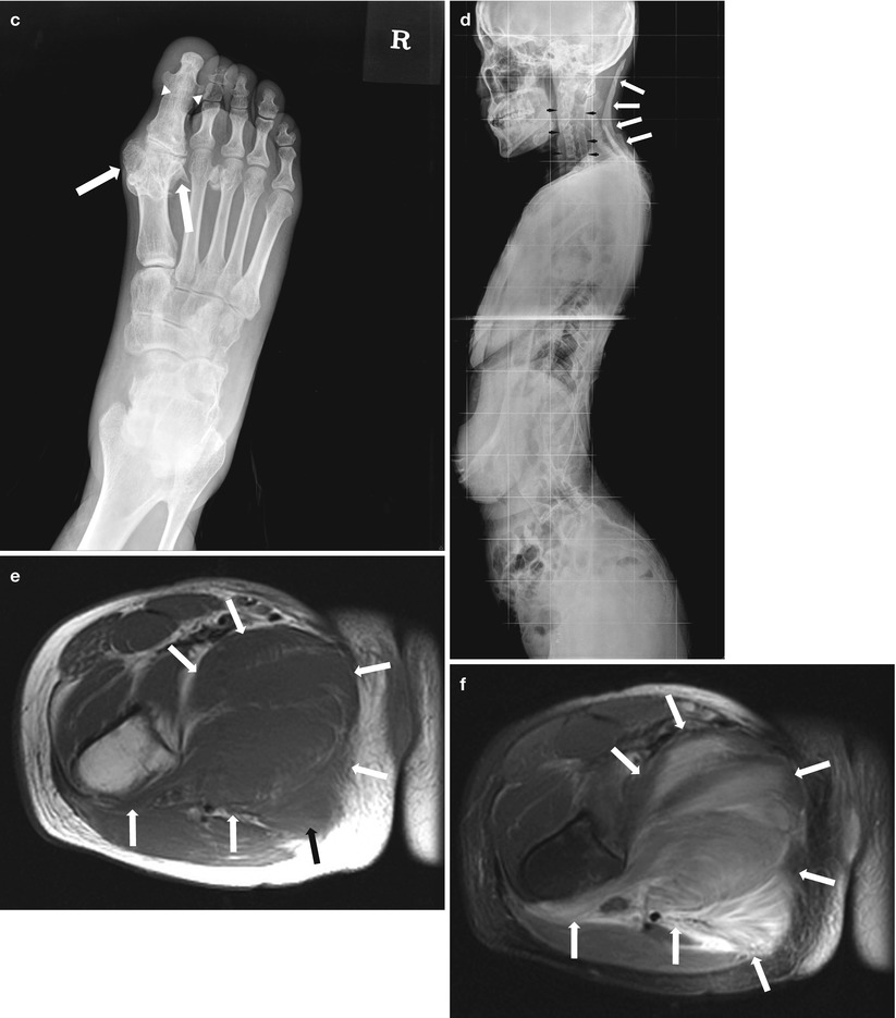
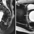
Fig. 29.1
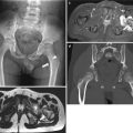
Radiographs and MR images of an 18-year-old girl who sustained myositis ossificans progressiva. She presented with edema of the right leg. (a) A radiograph of the left femur shows dense bridging ossification (arrows) in medial aspect of thigh. (b) Lateral radiograph of the elbow shows bony bridge (arrows) between the ventral aspect of humerus and ulna. (c) An anteroposterior (AP) radiograph of a foot shows dense soft tissue ossification resulting in the appearance of exostoses from the metatarsal bone of great toe with mild hallux valgus (arrows). Ankylosis of interphalangeal joint of great toe is also observed (arrowheads). (d) Lateral radiograph of the neck shows ankylosis of vertebral bodies and fact joints mimicking morphology of ankylosing spondylitis or Klippel-Feil syndrome (arrowheads). Note the bridging ossification bar (arrows) of the posterior neck portion presumably located at the nuchal ligament. (e) Axial T1-weighted MR image shows isointense signal intensity of the swollen muscles (arrows). (f) Axial T2-weighted fat-suppressed MR image shows hyperintense signal intensity of the swollen muscles at the medial and posterior compartments (arrows)
29.4.2 Infantile Hemangioma
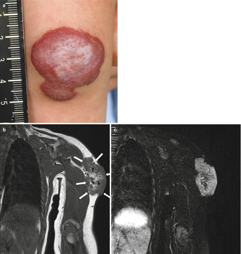
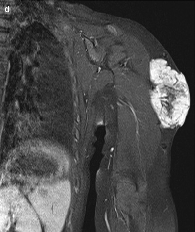
Fig. 29.2
Gross photograph and MR images of an 11-month-old girl with infantile hemangioma at the left arm. (a) Gross photograph shows a superficially located strawberry-like lump. (b) Coronal T1-weighted MR image shows well-defined isointense soft tissue mass (arrows) relative to muscle in subcutaneous through cutaneous layer of the Lt. arm. Intralesional high signal intensity portions (arrowheads) suggest imbibitions of fat component. (c) Coronal fat-suppressed T2-weighted MR image shows hyperintense lobulated soft tissue mass. (d) Coronal gadolinium-enhanced fat-suppressed T1-weighted MR image shows diffuse enhancement of the mass. Aforementioned intralesional fat component is not enhanced (Courtesy of In-One Kim and Young-Hun Choi of Seoul National University Hospital)
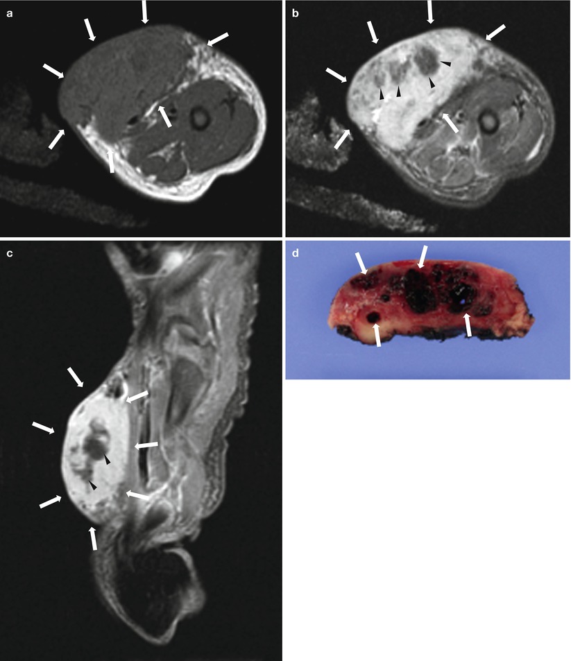
Fig. 29.3
MR images and gross photograph of a 4-day-old girl with infantile hemangioma at the left thigh. (a) Axial T1-weighted MR image shows a lobulated soft tissue mass (arrows) with isointense signal intensity relative to muscle confined to subcutaneous soft tissue of thigh. (b) Axial fat-suppressed T2-weighted MR image shows hyperintense lobulated soft tissue mass (arrows). Intralesional hypointense foci (arrowheads) reflect thrombi. (c) Sagittal gadolinium-enhanced fat-suppressed T1-weighted MR image shows diffuse enhancement of the mass (arrows). Nonenhanced foci (arrowhead) also reflect the presence of thrombi. (d) Photograph of the cut surface of the excised specimen shows multiple hemorrhagic cystic spaces (arrows) with/without thrombi (Courtesy of In-One Kim and Young-Hun Choi of Seoul National University Hospital)
29.4.3 Venous Malformation
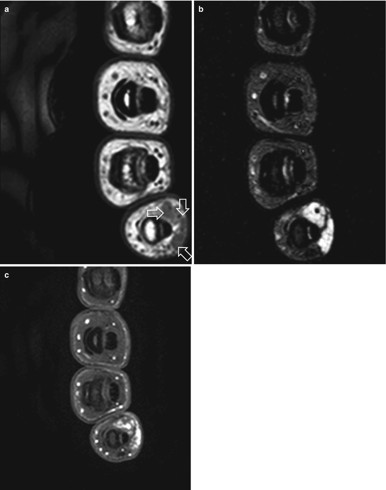
Fig. 29.4
MR images of an 18-year-old girl with venous malformation in the fifth finger. (a) Axial T1-weighted MR image shows well-defined soft tissue mass (arrows) which shows hypointense signal in volar aspect of fifth finger. (b) Axial fat-suppressed T2-weighted MR image of the mass shows a lobulated contour with multilocular appearance due to venous space that shows hyperintense signal separated by thin hypointense septa (arrowheads). (c) Axial gadolinium-enhanced fat-suppressed T1-weighted MR image of the mass shows patch enhancement
29.4.4 Lymphatic Malformation
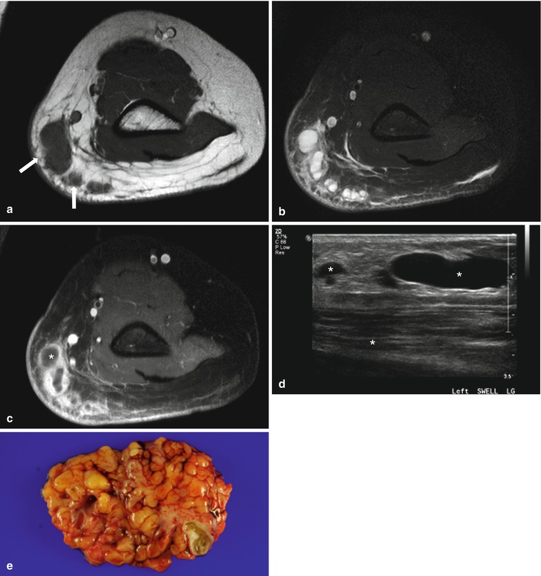
Fig. 29.5
MR images and gross photograph of a 13-year-old boy with lymphatic malformation at the left upper arm. (a) Axial T1-weighted MR image shows multiloculated subcutaneous mass (arrows) that is predominantly isointense relative to muscle, with some hyperintense subcutaneous fat interspersed within the lesion. (b) Axial fat-suppressed T2-weighted MR image shows the mass with multilocular cystic portions that show fluid signal. (c) Axial gadolinium-enhanced fat-suppressed T1-weighted MR image shows enhancement of surrounding connective tissue. No enhancement in central portion of cysts . Lack of enhancement of the lymph-filled space helps differentiated lymphatic malformation from venous malformation. (d) US shows multicystic fluid-filled mass in subcutaneous fatty layer of upper arm. (e) Gross photograph of completely excised specimen shows a multilobulated surface
29.4.5 Arteriovenous Malformation
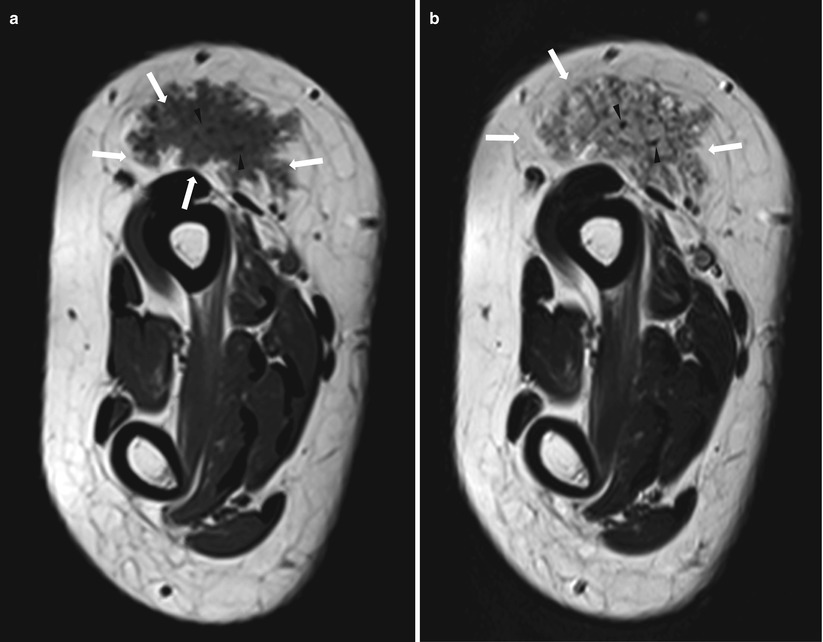
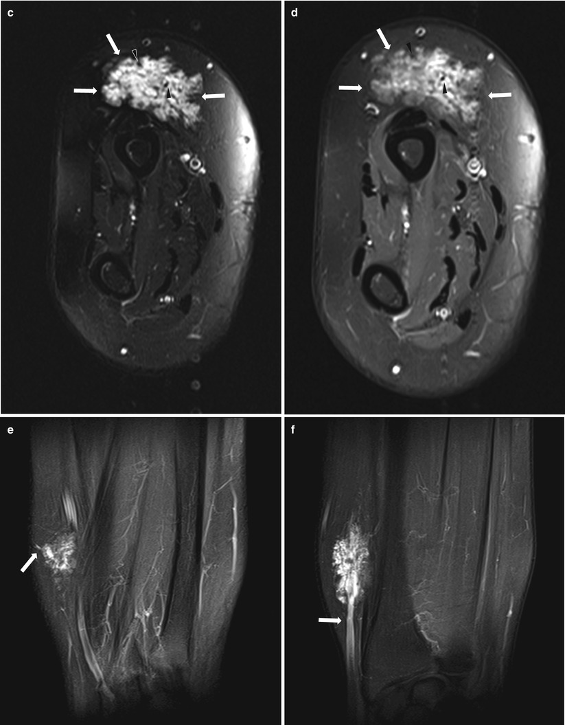
Fig. 29.6
MR images of a 17-year-old female patient with arteriovenous malformation. (a) Axial T1-weighted MR image shows isointense tangled mass (arrows) in subcutaneous layer of distal forearm. (b) Axial T2-weighted MR image shows the mass (arrows) with slightly hyperintense signal intensity relative to muscle. (c) Axial T2-weighted fat-suppressed MR image shows the mass (arrows) with bright signal intensity. (d) Axial gadolinium-enhanced fat-suppressed T1-weighted MR image shows heterogeneous enhancement of the mass (arrows). Note multiple signal void foci (arrowheads of a, b, c, and d) representing the high-flow vessels that are characteristic of this vascular malformation (e, f). Coronal gadolinium-enhanced fat-suppressed T1-weighted MR images show the supplying radial arterial branch (e, arrow) and engorged draining vein (f, arrow)
29.4.6 Klippel-Trenaunay Syndrome
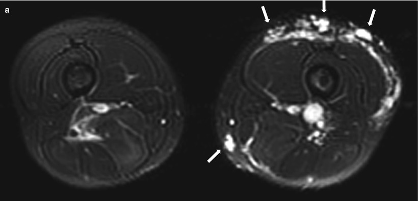
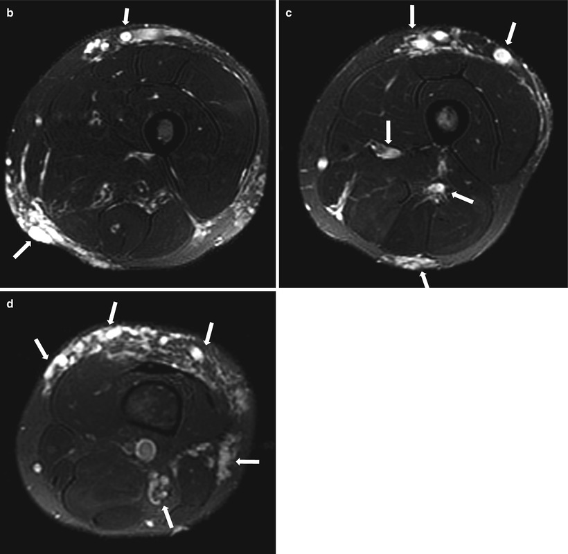
Fig. 29.7
MR images of a 19-year-old male patient who presented with asymmetric hypertrophy of the left thigh and is diagnosed as having Klippel-Trenaunay syndrome. (a) Axial fat-suppressed T2-weighted MR image shows asymmetric surface vein dilatation and vascular malformation (arrows) of the left thigh. Axial fat-suppressed T2-weighted MR image of upper (b), mid (c), and lower (d) level of the left thigh shows dilated vascular structures with vascular malformation involving subcutaneous and inter-/intramuscular area
29.4.7 Compartment Syndrome
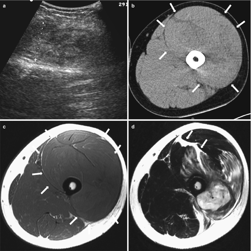
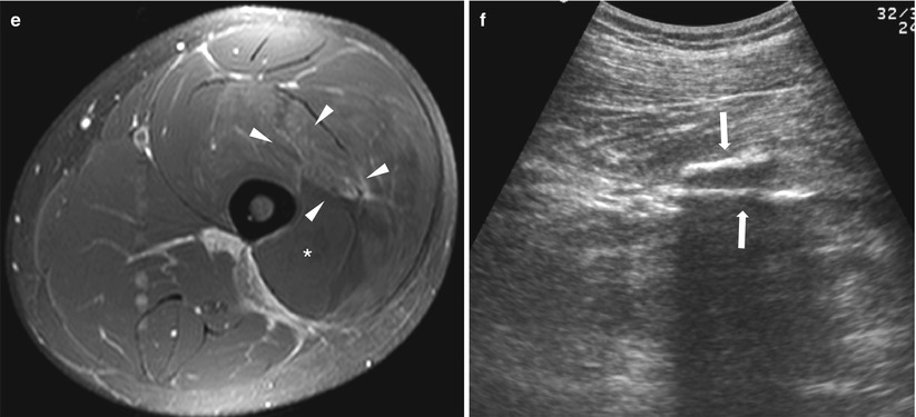
Fig. 29.8
Acute anterior compartment syndrome of the left thigh in an 18-year-old patient with swelling and pain after twisting injury. (a) US of left thigh shows diffuse swelling of vastus muscles with heterogeneous echo. (b) Precontrast CT scan also shows diffuse swelling of vastus muscles (arrows). (c) Axial T1-weighted MR image shows swelling of the entire anterior compartment muscles (arrows). (d) Axial T2-weighted MR image can reveal areas of muscle edema and a presumable hematoma within vastus muscles with hemorrhagic fluid (arrows) along the intermuscular facial planes. (e) Vastus intermedius and lateralis muscles show mild, patchy enhancement (arrowheads) on axial gadolinium-enhanced T1-weighted image. Note nonenhanced muscle area which was mentioned above as a hematoma. (f) Follow-up US after 2 months shows improved muscle edema with linear calcifications (arrows)
29.4.8 Fibromatosis Colli
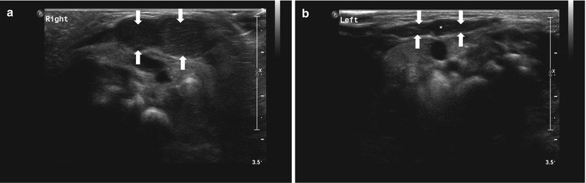
Fig. 29.9
US images of an 18-day-old boy who presented with fibromatosis colli. (a) Longitudinal oblique US image of the anterolateral neck shows nodular thickening of the sternocleidomastoid muscle (arrowheads). (b) Longitudinal oblique US image of the contralateral sternocleidomastoid muscle shows normal shape (arrows)
29.4.9 Desmoid Type Fibromatosis
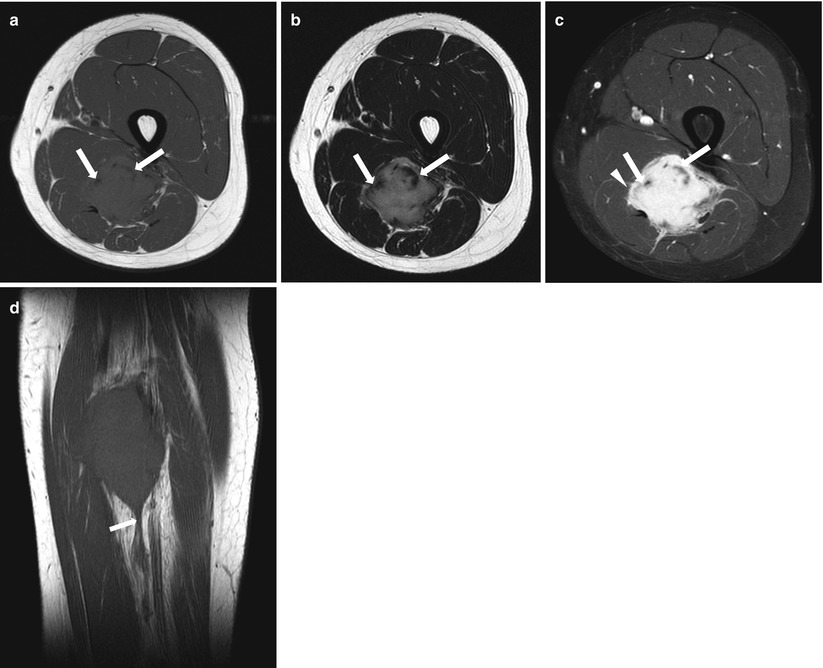
Fig. 29.10


MR images of a 12-year-old boy who sustained desmoid-type fibromatosis in the thigh. Three MR images at the same level (a, b, c) show a mass with lobulating border in posterior compartment of thigh. Axial T1-weighted (a) and axial T2-weighted (b) MR images show low signal intensity foci (arrow), which are more prominent on T2-weighted image. (c) Axial gadolinium-enhanced fat-suppressed T1-weighted MR image shows intense enhancement of the mass with infiltrative margins (arrowhead). The low signal foci seen on T1- and T2-weighted images are not enhanced partly (arrow), which suggest that the region is composed of collagen fibers. (d) Coronal T1-weighted MR image shows linear inferior extension of the mass along the fascia, which is called fascial tail sign (arrow)
Related posts:


Stay updated, free articles. Join our Telegram channel
Full access? Get Clinical Tree
