Solitary and Multiple Nodules, Masses, Cavities, and Cysts


Solitary and Multiple Nodules, Masses, Cavities, and Cysts


W. Richard Webb


The radiographic assessment of patients with solitary or multiple lung nodules, masses, or cavities is a common clinical problem. A primary or metastatic tumor usually is the first consideration in patients with these findings. However, many other diseases or abnormalities may present with focal lung abnormalities. Some have specific appearances that may suggest the correct diagnosis or limit the differential diagnosis.


THE SOLITARY PULMONARY NODULE

A solitary pulmonary nodule (SPN) usually is defined as a focal opacity, visible on chest radiographs or computed tomography (CT), which fits the following criteria:

1. It is relatively well-defined.

2. It is surrounded, at least partially, by lung.

3. It is roughly spherical in shape.

4. It is 3 cm or less in diameter (Fig. 9-1).

Lesions larger than 3 cm in diameter usually are referred to using the term “mass.” This cutoff also is used to distinguish a T1 carcinoma (3 cm or less in diameter) from a T2 carcinoma (more than 3 cm).


Clinical Evaluation

Clinical and historical information is helpful in the differential diagnosis of a solitary lung nodule. Important considerations that increase the likelihood of cancer include a history of smoking, age over 40, occupational exposures (e.g., asbestos), lung fibrosis, coexisting chronic obstructive pulmonary disease (COPD) and emphysema, and a family history of lung cancer. Risk factors for cancer are reviewed in Chapter 3.

Recent travel history, a positive skin test for tuberculosis (TB) or fungus, or the presence of other diseases (e.g., rheumatoid arthritis) increases the likelihood of a benign SPN. In a patient younger than 30 years of age, cancer is very uncommon as a cause of SPN.

An SPN found in a patient with a history of extrathoracic malignancy may be a metastasis, a primary lung carcinoma, or an insignificant benign lesion. Certain tumors have a propensity to metastasize as SPNs. Among patients with an SPN, those with a history of melanoma, sarcoma, or testicular carcinoma are more than twice as likely to have a solitary metastasis than a bronchogenic carcinoma. On the other hand, patients with an SPN who have a history of carcinoma of the head and neck, bladder, breast, cervix, bile ducts, esophagus, ovary, prostate, or stomach are more than three times as likely to have primary bronchogenic carcinoma than lung metastasis. In patients with other types of tumors, the relative likelihood of metastasis and lung cancer is about equal.


Radiographic Evaluation

The differential diagnosis of an SPN is extensive (Table 9-1). In patients with an SPN, plain films and CT are used to determine the nodule’s (1) morphologic characteristics, (2) density or attenuation (i.e., calcium, fat, or contrast enhancement), and (3) growth rate.


Morphologic Characteristics

Using plain radiographs or CT, an SPN sometimes may be diagnosed as a specific lesion based on its appearance. Some such lesions include mucous plug, arteriovenous fistula, rounded atelectasis, mycetoma, and focal pleural lesions. These are described later in this chapter.

Much more often, radiographic studies are used to assess less specific morphologic characteristics that suggest that an SPN is likely malignant or likely benign.


Size

The likelihood of malignancy in a nodule or mass is directly related to its size (Table 9-2). The likelihood of cancer is about 35% for a nodule ranging from 0.5 to 1.0 cm in diameter, 50% for an SPN 1.0 to 2.0 cm in diameter, and more than 85% for an SPN more than 2.0 cm in diameter (see Fig. 9-1). It should be kept in mind, however, that even a very small lesion can represent a carcinoma.

One or more small lung nodules are exceedingly common as an incidental finding on CT scans, and the large majority represent benign lesions such as granuloma or intrapulmonary lymph node. Follow-up CT is the only practical method of evaluating their significance.


FIG. 9.1. Right upper lobe nodule representing an adenocarcinoma. A: Chest radiograph shows a right upper lobe nodule (arrow). This lesion is classified as a nodule because it is relatively well-defined, at least partially surrounded by lung, roughly spherical in shape, and 3 cm or less in diameter. Because the nodule exceeds 2 cm in diameter, it is very likely malignant. B: CT shows the edge of the nodule to be ill defined and lobulated, and a pleural tail (arrow) extends to the pleural surface.


TABLE 9.1 Differential Diagnosis of an SPN or Mass


Congenital Lesions and Normal Variants


Arteriovenous fistula


Bronchogenic cyst


Congenital cystic adenomatoid malformation


Intrapulmonary lymph node


Mucoid impaction (bronchial atresia)


Pulmonary vein varix


Sequestration


Malignant Neoplasms


Carcinoma


Lymphoma


Lymphoproliferative disease


Metastatic neoplasm


Sarcoma (e.g., chondrosarcoma, liposarcoma, fibrosarcoma)


Benign Neoplasms and Neoplasm-like Conditions


Endometrioma


Hamartoma


Lymphoproliferative disease


Miscellaneous benign tumors


Mesenchymal tumors (e.g., chondroma, lipoma, fibroma)


Epithelial tumors (e.g., atypical adenomatous hyperplasia, mucous gland adenoma)


Vascular tumors


Infection and Parasites


Aspergillosis, angioinvasive


Dirofilaria immitis (dog heartworm)


Echinococcus


Focal (round) pneumonia


Granulomatous infection or granuloma


Tuberculosis


Nontuberculous mycobacteria (e.g., Mycobacterium avium-intracellulare complex)


Coccidioidomycosis


Histoplasmosis


Cryptococcus


Lung abscess


Mycetoma (aspergilloma)


Pulmonary gangrene


Septic embolism


Inflammatory (Noninfectious)


Churg-Strauss syndrome


Focal organizing pneumonia


Rheumatoid nodule


Sarcoidosis


Wegener’s granulomatosis


Airways and Inhalational Disease


Mucoid impaction (mucous plug)


Asthma


Allergic bronchopulmonary aspergillosis


Bronchial atresia


Bronchiectasis


Cystic fibrosis


Conglomerate mass or progressive massive fibrosis (e.g., silicosis)


Lipoid pneumonia


Vascular Lesions


Arteriovenous fistula


Hematoma


Infarction


Pulmonary artery aneurysm


Pulmonary vein varix


Septic embolism


Idiopathic and Miscellaneous


Amyloidosis


Fluid-filled bulla


Round atelectasis


TABLE 9.2 Likelihood of Malignancy Related to Nodule Diameter


Diameter (cm)


Malignancy Rate (%)


<1


35


1-2


50


2-3


80


>3


97


Location

About two thirds of lung cancers occur in the upper lobes, and the right upper lobe is most commonly involved (see Fig. 9-1). Sixty percent of cancers presenting as an SPN on chest radiographs are seen in the lung periphery; only 10% are visible in the medial third of the lung.

A metastatic tumor presenting as an SPN tends to be located in the subpleural or outer third of the lung. Two thirds of metastatic lesions occur in the lower lobes.


Edge Appearance

Although plain radiographs do not allow the edge of a lung nodule or mass to be assessed with the precision of CT, cancers can appear to be ill defined, irregular in contour, spiculated, or lobulated on plain films (see Fig. 9-1).


FIG. 9.2. Adenocarcinoma with a spiculated margin seen on CT. Two pleural tails (arrows) extend to the pleural surface. This appearance has been termed corona radiata or corona maligna. The surface of the nodule is lobulated and shows notches, both of which are findings indicating malignancy. Several lucencies within the nodule represent air bronchograms of areas of pseudocavitation, typical findings in adenocarcinoma and BAC.


FIG. 9.3. Smooth, sharply marginated, rounded nodule representing a granuloma. Its appearance (arrow) is typical of a benign lesion. Its small size also makes malignancy less likely.

On CT, malignant nodules are much more likely to have an ill-defined, irregular, lobulated, or spiculated margin (see Figs. 9-1B and 9-2). Benign lesions tend to have a smooth, sharply defined edge (Figs. 9-3 and 9-4; Table 9-3). Nearly 90% of nodules with an irregular or spiculated edge are malignant; only 20% of nodules with a smooth, sharp margin are malignant. Malignancies that tend to have a sharp and smooth edge include metastases (Fig. 9-5) and carcinoid tumors (see Fig. 3-44 in Chapter 3).

The terms corona radiate and corona maligna have been used to describe the appearance of spiculation associated with a nodule or mass (see Figs. 9-2 and 9-6). Particularly in patients with adenocarcinoma and bronchioloalveolar carcinoma (BAC), this appearance reflects the presence of fibrosis surrounding the tumor, although tumor invasion of the adjacent lung also may be present. The fibrosis usually reflects a desmoplastic reaction rather than preexisting lung fibrosis. Spiculation is less common with large cell carcinoma than other cell types that present as a solitary nodule or mass.

In addition, carcinomas can show the presence of a pleural tail sign, in which a thin linear opacity is seen extending from the edge of a lung nodule to the pleural surface (see Figs. 9-1B, 9-2, and 9-6). This tail, which can be from a few millimeters to a few centimeters in length, often is seen in association with spiculation. As with spiculation, it reflects the presence of fibrosis and often is associated with a dimpling of the visceral pleura. In patients with lung cancer, a pleural tail sign most often is associated with adenocarcinoma or BAC; it uncommonly indicates the presence of a large cell carcinoma. The pleural tail sign also can be seen in
association with benign lung nodules, which are associated with fibrosis, including various granulomatous diseases. The presence of a spiculated contour is more suggestive of malignancy than a pleural tail.


FIG. 9.4. Hamartoma presenting as a sharply defined, round nodule. A: Chest radiograph shows a round nodule (arrows) in the right upper lobe. B: CT shows the nodule (arrow) to be rounded in shape and sharply marginated. Slight lobulation may be seen with hamartomas.

The halo sign, a halo of ground-glass opacity surrounding a lung nodule, may be seen in some patients with an SPN. It is commonly present in leukemic patients with angioinvasive aspergillosis (Fig. 9-7) but also can be seen in patients with other infections (see Fig. 9-35C) and in some tumors, particularly adenocarcinoma or BAC (Fig. 9-8; Table 9-4). The histologic nature of the halo varies with the disease. In patients with invasive aspergillosis, the halo sign represents hemorrhage; in patients with carcinoma, it reflects the presence of lepidic spread of tumor.


TABLE 9.3 Edge Appearances and Common Diagnoses


Sharply marginated


Granuloma


Hamartoma or benign tumor


Carcinoid tumor


Metastasis


Spiculated (corona radiata) or pleural tail


Bronchioloalveolar carcinoma


Carcinoma


Granuloma or focal scarring


FIG. 9.5. Solitary metastasis from a head and neck carcinoma. A left upper lobe nodule (arrow) is smooth and sharply defined on CT. This appearance is common with metastases.


FIG. 9.6. Adenocarcinoma. HRCT shows an irregular, spiculated nodule with multiple pleural tails. Air bronchograms are visible within the nodule.


Shape

Lung carcinomas tend to be irregular in shape, lobulated, or notched (see Figs. 9-1, 9-2, and 9-6) but uncommonly have concave margins. Granulomas often are round (see Fig. 9-3). Hamartomas and metastases may be round, oval, or lobulated (see Figs. 9-4 and 9-5). Scars or areas of atelectasis or scarring may appear linear or angular. A number of other benign lesions (e.g., AVM, mucous plugs) may be identified by their characteristic shapes.


FIG. 9.7. Halo sign in invasive aspergillosis. HRCT in a young patient with leukemia and granulocytopenia shows a dense left lower lobe nodule surrounded by a halo (arrows) of ground-glass opacity. In patients with invasive aspergillosis, the halo represents hemorrhage surrounding a septic infarction.


FIG. 9.8. Halo sign in BAC. HRCT shows a dense central nodule surrounded by a halo (arrows). In BAC, the halo represents the presence of lepidic tumor growth.


Air Bronchograms and Pseudocavitation

On HRCT, air bronchograms commonly are seen in cancers presenting as an SPN (25% to 65% of cases; see Figs. 9-2 and 9-6). This finding is most typical of adenocarcinoma or BAC. Air bronchograms are much less common in benign lesions but have a variety of causes, such as focal pneumonia, infarction, round atelectasis, or conglomerate masses in patients with silicosis or sarcoidosis (Table 9-5). An appearance mimicking the presence of air bronchograms may be seen in patients with developing mycetoma; the apparent air bronchograms
represent air-filled spaces between fronds of fungus. Small bronchi seen in relation to lung cancers often appear abnormal, being narrowed, obstructed, or irregular in contour.


TABLE 9.4 Causes of the Halo Sign


Fungi: invasive aspergillosis, candidiasis, coccidioidomycosis


Bacteria: tuberculosis, Nocardia, Legionella


Viruses: cytomegalovirus, herpes


Pneumocystis jiroveci (P. carinii)


Organizing pneumonia


Wegener’s granulomatosis


Infarct


Metastatic tumor


Kaposi’s sarcoma


Bronchioloalveolar carcinoma


Adenocarcinoma


TABLE 9.5 Causes of Air Bronchograms in SPNs


Adenocarcinoma


Bronchioloalveolar carcinoma


Conglomerate mass (e.g., silicosis, sarcoidosis)


Focal pneumonia


Infarction


Rounded atelectasis


Bronchiolitis obliterans with organizing pneumonia


Lymphoma


Lymphoproliferative diseases


Mycetoma (may mimic an air bronchogram)


In addition to air bronchograms, small bubbly lucencies may be seen in cancers (see Fig. 9-2). These may represent air bronchograms, small air-filled cystic areas in the tumor (so-called pseudocavitation), or small cavities. They have the same significance as air bronchograms.


Cavitation

By general agreement, a cyst is an air-filled lesion that has a smooth and uniform wall 3 mm or less in thickness. The term cavity usually is used to describe a lesion with a thicker or more irregular wall or a lesion that has cavitated (i.e., evolved by developing an air-filled space, regardless of how thick the wall is). Thus, a thin-walled lesion may be either a cyst or a cavity, whereas a thick-walled or irregular lesion is a cavity. An exception to this rule is an infected cyst: surrounding lung inflammation may result in a thick “wall.”


FIG. 9.9. Cavitary carcinoma. A: Plain radiograph showing a cavitary left lung mass that represents a squamous cell carcinoma. B: Cavitary squamous cell carcinoma shown at two levels. The wall of the cavity is irregular, with several thick nodular regions (white arrow). The cavity contains an air-fluid level (black arrows). This is uncommon in malignancy and may represent hemorrhage or infection. C: Cavitary adenocarcinoma shown on HRCT in six contiguous scans. The nodule contains an irregular cavity; is irregular and lobulated in shape, notched, and spiculated; and is associated with pleural tails. It also contains several air bronchograms.

Cavitation occurs in about 10% of cancers, most commonly in patients with squamous cell carcinoma (Fig. 9-9A and B; see also Figs. 3-4 and 3-26 in Chapter 3). Approximately 80% of cavitary lung cancers are squamous cell carcinomas. Cavitation of large cell carcinoma and adenocarcinoma also occurs (see Fig. 9-9C); small cell carcinoma rarely cavitates.

Although a long list of abnormalities may be associated with cysts or cavities, described in Table 9-6 and in detail below, for practical purposes, radiographic evaluation is directed at determining the likelihood of malignancy. Cavitary malignant lesions tend to have a thick, nodular wall (see Fig. 9-9B and C; see also Figs. 3-4 and 3-26 in Chapter 3); benign lesions often have a thin, smooth wall (Fig. 9-10). The thickness of the wall of a cavity serves as an indicator of its likelihood of being malignant. Nearly 85% of cavities with a wall measuring more than 15 mm in its thickest portion are malignant (see Fig. 9-9B). If the thickest part of the wall is less than 5 mm, 95% are benign (see Fig. 9-10). Seventy-five
percent of cavities with a wall 5 to 15 mm in thickness are benign. If the thickest part of the cavity wall measures 1 mm or less, malignancy is rare. However, thin-walled cystic lesions may rarely be seen with BAC or metastases.


TABLE 9.6 Causes of a Cyst or Cavity (Solitary or Multiple)


Amyloidosis—solitary or multiple


Aspergillosis, angioinvasive—usually multiple


Aspergillosis, semi-invasive—usually solitary


Bronchogenic cyst—usually single


Bulla—solitary or multiple


Carcinoma


Congenital cystic adenomatoid malformation—solitary, but often multiloculated


Conglomerate mass or progressive massive fibrosis—often bilateral


Cystic bronchiectasis—usually multiple


Cystic lung disease (e.g., histiocytosis, lymphangiomyomatosis)—multiple


Echinococcus—solitary or multiple


Endometrioma


Granulomatous infection


Tuberculosis


Nontuberculous mycobacteria (e.g., Mycobacterium avium-intracellulare complex)


Coccidioidomycosis


Histoplasmosis


Cryptococcus


Hematoma—solitary or multiple


Intralobar sequestration—may be lucent, cystic, or multicystic


Lung abscess—solitary or multiple


Lymphoma—solitary or multiple


Metastatic neoplasm—usually multiple


Mycetoma—aspergilloma


Papillomatosis—usually multiple


Paragonimiasis—usually multiple


Pneumatocele—solitary or multiple


Pulmonary gangrene—usually solitary


Pulmonary laceration—traumatic


Rheumatoid nodule—usually multiple


Sarcoidosis—usually multiple


Sarcoma—solitary


Septic embolism—usually multiple


Wegener’s granulomatosis—usually multiple


Lung cancer resulting in bronchial obstruction also can be associated with an abscess in the distal lung, mimicking a cavitary carcinoma. In addition, lung cancer sometimes can arise in a bulla or cyst or be associated with a preexisting cavity. In such a case, focal thickening of the cyst or cavity wall or fluid within the cyst may be the only findings suggesting this diagnosis.


Air-crescent Sign

In some patients with a cavitary nodule or lung cyst, a mass or nodule may be present within the cavity. Air outlining or capping the superior aspect of the mass results in a crescent-shaped collection of air, termed the “air-crescent” sign. The most likely cause of this appearance is aspergilloma (mycetoma), but the differential diagnosis includes other entities as well (Table 9-7). Gravitational shift of the intracavity mass strongly suggests mycetoma and excludes carcinoma.


FIG. 9.10. CT scan of a lung abscess. The wall is thin and smooth, measuring less than 5 mm in thickness. An air-fluid level is visible.


Air-fluid Level

The presence of an air-fluid level in a patient with a cavitary SPN tends to indicate a benign lesion, particularly lung abscess (see Fig. 9-10). Any infected cystic or cavitary lesion may be associated with an air-fluid level. An air-fluid level is
uncommon in a cavitary carcinoma but may be seen in the presence of hemorrhage or superinfection (see Fig. 9-9B).


TABLE 9.7 Causes of the Air-crescent Sign


Aspergilloma (mycetoma)


Angioinvasive aspergillosis with septic infarction


Carcinoma arising in a cyst


Cavitary carcinoma


Clot in a cyst or cavity


Echinococcus


Mucous plug in cystic bronchiectasis


Papillomatosis


Pulmonary gangrene


Rasmussen aneurysm (mycotic pulmonary artery aneurysm in a tubercular cavity)


FIG. 9.11. Tuberculosis. A right upper lobe nodule is associated with satellites (arrows). This appearance is most typical of a benign process but sometimes is seen with carcinoma.


Satellite Nodules

Satellite nodules are small nodules seen adjacent to a larger nodule or mass. They tend to predict a benign lesion (Fig. 9-11). Satellites are most common with granulomatous diseases and infections such as TB (Table 9-8). Only a small percentage of carcinomas are associated with satellite nodules. In patients with sarcoidosis, the presence of satellite nodules has been termed the “galaxy” sign.


Feeding Vessel Sign

The “feeding vessel” sign is present if a small pulmonary artery is seen leading directly to a nodule (Fig. 9-12). This appearance is most common with metastasis, infarct, and arteriovenous fistula. It is less common with primary lung carcinomas or benign lesions such as granuloma.


TABLE 9.8 Causes of Satellite Nodules


Tuberculosis


Nontuberculous mycobacterial infection


Bacterial infections with endobronchial spread


Fungal infections


Sarcoid


Conglomerate masses (silicosis, coal worker’s pneumonoconiosis, talcosis)


Bronchioloalveolar carcinoma


Adenocarcinoma


FIG. 9.12. Metastatic nasopharyngeal carcinoma. Multiple nodules (arrows) are associated with a feeding vessel.


Attenuation

A lung nodule usually is examined using volumetric HRCT to determine its attenuation before contrast injection. Because of volume averaging, unless an SPN is grossly calcified, CT with thick collimation (5 mm) usually cannot be used to determine its attenuation accurately. Most cancers appear to be of soft tissue attenuation.


Ground-glass Opacity

With thin collimation, some nodules appear to be of ground-glass opacity. Many focal opacities of ground-glass opacity are inflammatory and resolve on follow-up. However, BAC may present as a nodule entirely of ground-glass opacity, and a high degree of suspicion should be maintained (Fig. 9-13). Follow-up of such a lesion is appropriate.


Calcification or High Attenuation

The presence of calcium in an SPN increases its chances of being benign (Table 9-9). Diagnosing a small nodule as calcified on chest radiographs is somewhat subjective and subject to error. However, if a nodule a few millimeters in diameter is easily seen on radiographs, it probably is calcified.


FIG. 9.13. Bronchioloalveolar carcinoma. A spiculated nodule (arrows) is visible on CT with 3-mm slice thickness. The nodule is of ground-glass opacity. This appearance may be seen with BAC.

CT is more sensitive and accurate in diagnosing calcification. Volumetric HRCT images usually should be obtained through a lung nodule to look for calcium. HRCT demonstrates calcification in about 25% of benign SPNs that do not appear calcified on plain radiographs.

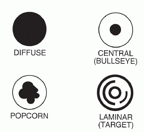
The pattern of calcification is important in determining its diagnostic significance. Generally, the following four patterns of calcification can be used to predict the presence of a benign lesion with sufficient accuracy to allow appropriate management (Fig. 9-14):


TABLE 9.9 Causes of Calcification or High Attenuation in an SPN


Amyloidosis—dense or stippled


Carcinoid tumor—punctuate, eccentric


Carcinoma—punctate, eccentric


Conglomerate mass—multiple foci


Dirofilaria immitis


Granuloma—diffuse, central, concentric


Hamartoma or chondroma—popcorn, central


Mucoid impaction in allergic bronchopulmonary aspergillosis or bronchial atresia


Metastases—diffuse, punctuate


Talcosis—secondary to talc not calcium


Amiodarone toxicity—due to iodine content


FIG. 9.14. Benign patterns of calcification. With rare exceptions, these indicate the presence of a benign lesion.


  • Homogeneous calcification (Fig. 9-15)


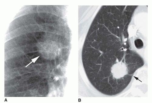
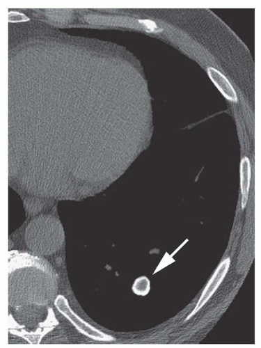
  • Dense central (bull’s-eye) calcification (Fig. 9-16)


  • Concentric rings of calcium (“target” calcification; Fig. 9-17)


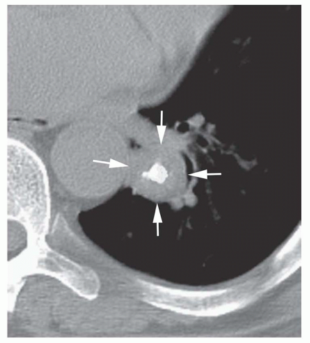
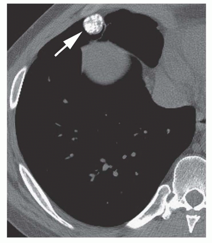
  • Conglomerate foci of calcification involving a large part of the nodule (“popcorn” calcification; Fig. 9-18)


FIG. 9.15. Homogeneous calcification. Dense and uniform calcification of a small right upper lobe nodule (arrow) is typical of a benign lesion, usually a tuberculoma.


FIG. 9.16. Dense central or “bull’s-eye” calcification in a hamartoma. A round lung nodule (arrows) adjacent to the descending aorta shows dense central calcification. This is typical of histoplasmoma or hamartoma.

The first three of these patterns are most typical of granulomas; the last is more typical of hamartoma. Calcified lesions thought to be benign should be followed up radiographically in most cases unless the calcification is diffuse.


FIG. 9.17. Concentric or “target” calcification (arrow). One or more rings of calcium may be seen. This pattern is typical of a histoplasmoma.


FIG. 9.18. Multiple confluent nodular foci of calcification (“popcorn” calcification; arrow) in a hamartoma. This appearance is typical of hamartoma and corresponds to the calcification of cartilage nodules.

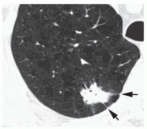
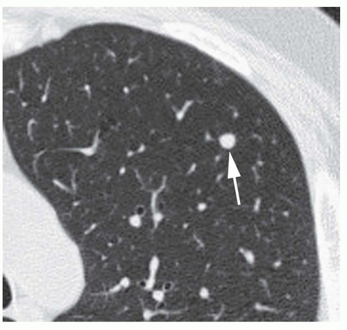
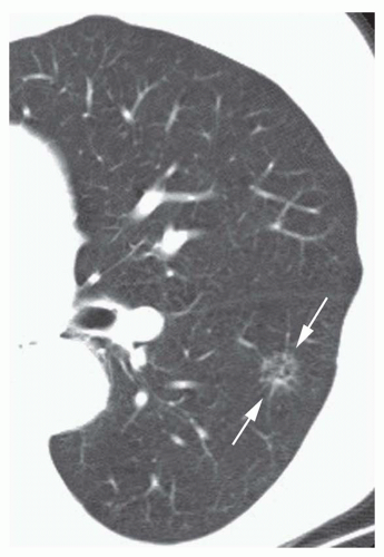
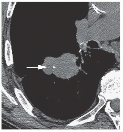
Stippled calcification or eccentric foci of calcification (Figs. 9-19 and 9-20) may be seen in benign SPNs but are visible in as many as 10% to 15% of cancers; these patterns must be considered indeterminate (see Fig. 9-19).

Calcium in a tumor may reflect dystrophic calcification (occurring in areas of tumor necrosis), engulfing of a preexisting granuloma, or calcification of the tumor itself (as in mucinous adenocarcinoma, carcinoid tumor, or osteogenic sarcoma). A “benign” pattern of calcification occasionally is seen in patients with neoplasm. Carcinoid tumor and mucinous adenocarcinoma can show dense central calcification. Metastases from osteogenic sarcoma or chondrosarcoma can show homogenous calcification, but a history of the primary tumor allows a correct diagnosis.

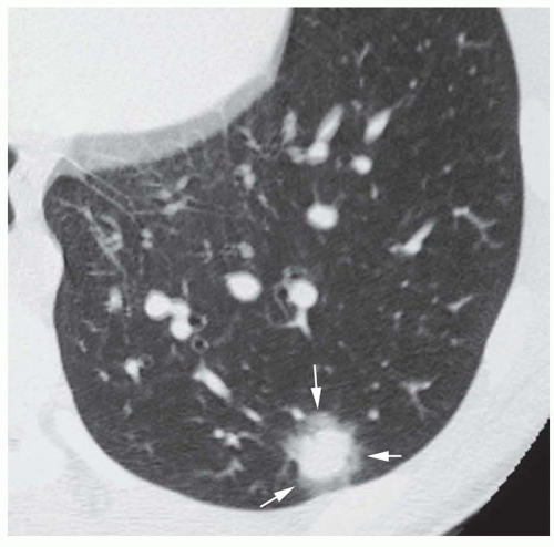
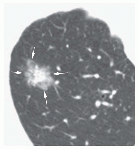
Because of the high likelihood of cancer in patients with spiculated nodules or nodules exceeding 2 cm in diameter, it is inadvisable to call such a nodule benign on the basis of visible calcification, unless the calcification is diffuse and dense. A small central calcification is insufficient for determining whether such a nodule is benign.

Usually, a visible inspection of HRCT scans is sufficient for diagnosing calcification. However, the measurement of
CT numbers can allow the detection of calcification, which is not clearly seen on the scans. This technique is termed CT nodule densitometry. Pixels denser than 100 HU indicate the presence of calcification.


FIG. 9.19. Indeterminate patterns of calcification. These may be seen in benign or malignant lesions.


FIG. 9.20. Eccentric calcification in an adenocarcinoma. A lobulated mass shows a small focus of eccentric calcification (arrow).

High attenuation of a nodule or mass sometimes is seen using CT in patients who do not have calcification. This may be seen in patients with amiodarone toxicity resulting in focal organizing pneumonia: the drug contains iodine, which appears dense. Patients with conglomerate masses from talcosis may show high attenuation due to the talc.
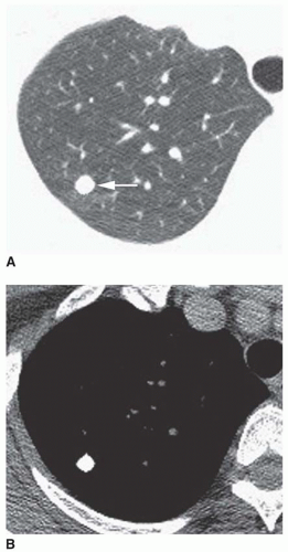

Fat

The presence of fat in an SPN may be diagnosed accurately only on HRCT. On HRCT, fat can be accurately diagnosed if low CT numbers are seen (-40 to -120 HU). This most likely indicates the presence of hamartoma (see Figs. 3-49 and 3-51 in Chapter 3), lipoma, or lipoid pneumonia (see Figs. 20-1 and 20-2 in Chapter 20). On occasion, histoplasmoma may show fat deposition with growth (
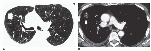
Table 9-10). Nearly 65% of hamartomas show fat on HRCT, sometimes in association with dense popcorn calcification or flecks of calcium. The presence of fat within a lung nodule is sufficient for calling it benign, although follow-up is appropriate.


TABLE 9.10 Causes of SPNs Containing Fat


Hamartoma


Lipoma


Liposarcoma (primary or metastatic)


Lipoid pneumonia


Histoplasmoma


Teratoma


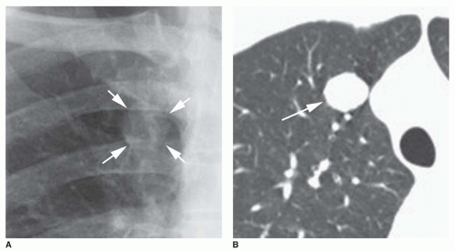
Low (Water or Fluid) Attenuation

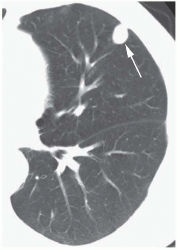
Benign cystic lesions, such as pulmonary bronchogenic cyst, sequestration, congenital cystic adenomatoid malformation (CCAM), or a fluid-filled cyst or bulla (Table 9-11), occasionally may be diagnosed on CT by their low attenuation (0 to 20 HU) and very thin or invisible walls (see Fig. 1-6 in Chapter 1). Mucoid impaction also may appear to be of low attenuation. On the other hand, bronchogenic cysts or other cystic lesions may have a higher attenuation because of their protein content. A hematoma may have the attenuation of blood (50 HU) or may have low attenuation, depending on its age.

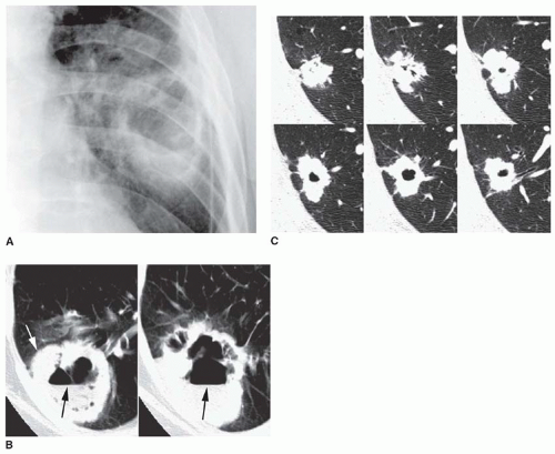
A necrotic neoplasm, conglomerate mass, or lung abscess or infarction may have a low attenuation center on CT, but these lesions have a thick and perceptible wall.


Contrast Enhancement

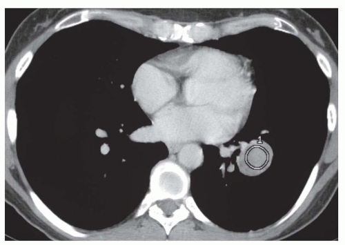
Cancers have a greater tendency to opacify following contrast infusion than do some types of benign nodules. Specific contrast enhancement techniques have been suggested to help diagnose malignancy. When using these techniques, sequential thin-collimation scans must be obtained through the center of a lung nodule for several minutes following contrast injection. Most SPNs show peak enhancement at either 3 or 4 minutes.

One currently recommended protocol uses scans at 1 minute intervals for 4 minutes following the start of the injection of 420 mg iodine/kg (usually 75 to 125 mL) at a rate of 2 mL/s. A region of interest encompassing about 60% of the nodule diameter is used to measure enhancement.

Using this protocol, carcinomas enhance by 14 to 165 HU (median, 38 HU; Fig. 9-21) and benign lesions exhibit a change in CT number of -20 to 96 HU (median, 10 HU). Using an enhancement of 15 HU or more to suggest
malignancy, this test has a sensitivity of 98%, specificity of 58%, and accuracy of 77%. Specific benign lesions that show significant enhancement include active granulomas, inflammatory lesions, focal pneumonias, and some benign tumors such as hamartoma (Fig. 9-22; Table 9-12). Round atelectasis tends to enhance densely, as does any area of atelectasis.


TABLE 9.11 Causes of Fluid Density SPNs


Bronchogenic cyst


Carcinoma (necrotic or infected)


Congenital cystic adenomatoid malformation


Conglomerate mass (necrotic)


Fluid-filled or infected cyst, cavity, or bulla


Hematoma


Lung abscess (bacterial and fungal)


Lymphoma (necrotic)


Metastatic neoplasm (necrotic)


Mucoid impaction


Sequestration


FIG. 9.21. Enhancement of a carcinoma. A: A lobulated right upper lobe nodule shown on HRCT represents a carcinoma. B: After contrast enhancement, the region of interest in the center of the nodule (arrow) showed an increase in attenuation of 40 HU (change from 8 to 48 HU). This is typical of malignant lesions.

It would seem most appropriate to use this technique when a nodule does not show typical findings of malignancy (e.g., spiculation, nodularity, cavitation, or growth) or typical features of a specific benign lesion. In such patients, this technique may help select which patients require surgery (i.e., if enhancement is present) or which can just be followed up carefully (i.e., if enhancement is absent).


FIG. 9.22. Enhancement of a hamartoma. HRCT through a left lung nodule during contrast enhancement showed an increase in attenuation of 22 HU (increase in attenuation from 20 to 42 HU) following contrast enhancement for the region of interest shown.


Contrast Opacification

Some solitary (or multiple) lesions opacify following contrast injection, thus representing vascular structures (Table 9-13). These have a limited differential diagnosis and specific morphology, described in the following sections.


Growth and Doubling Time

Carcinomas grow (Fig. 9-23). The growth rate of an SPN has been used to determine its likelihood of being malignant. Doubling time, the time required for a lesion to double in volume, is used to measure the growth rate. For easy reference, a 26% increase in nodule diameter is one doubling, and a doubling of diameter means that three volume doublings have occurred. However, not all carcinomas grow in a concentric fashion, and estimating their volume may be difficult (see Fig. 9-23).

Only gold members can continue reading. Log In or Register to continue

Stay updated, free articles. Join our Telegram channel

Oct 10, 2016 | Posted by in CARDIOVASCULAR IMAGING | Comments Off on Solitary and Multiple Nodules, Masses, Cavities, and Cysts

Full access? Get Clinical Tree

Get Clinical Tree app for offline access