Chapter 20 Specialised projections of the skull
Sella turcica (pituitary fossa)
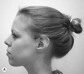
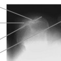
Lateral sella turcica (Fig. 20.1A,B)
Positioning
• The patient is initially seated facing the IR
• The trunk is brought as close as possible to the receptor unit and the patient is asked to sit with their spine as erect as possible. This helps the patient turn their head more easily into the required lateral position
• The head is turned through 90° to bring the side of the head in contact with the IR
• The median sagittal plane (MSP) is parallel to the IR; there should be no tilt or rotation of the head. This can be assessed by checking the midline of the cranium over the top and symmetry of the frontal bone and orbits

Criteria for assessing image quality
• Anterior and posterior clinoid processes, sella turcica, sphenoid sinus and dorsum sellae are demonstrated
• Superimposition of both sides of the floor of pituitary fossa and floor of the anterior cranial fossa
• Anterior clinoid processes are superimposed
• Posterior clinoid processes are superimposed
• Sharp image demonstrating outline of the clinoids and pituitary fossa in contrast with the temporal bones and sphenoid sinus
| Common errors | Possible reasons |
|---|---|
| Both pairs of clinoid processes overlapped, seen one above the other | Tilted skull |
| Both pairs of clinoid processes overlapped, seen side by side | Rotated skull |
| ’Double’ floor of sella turcica; two lines over floor area | Tilted skull or floor is eroded on one side by tumour |
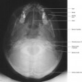
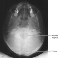
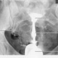
Occipitofrontal (OF) sella turcica (Fig. 20.2A,B,C)
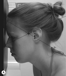
Beam direction and FRD
(a) Initially horizontal, a 20° caudal angle will demonstrate the floor of the pituitary fossa through the ethmoid and sphenoid sinuses
(b) A 30° cranial angle will demonstrate the dorsum sellae through the foramen magnum
Criteria for assessing image quality
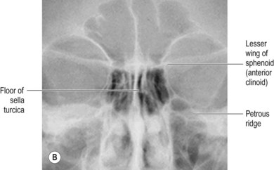
OF 20°
• Lesser wing of the sphenoid, sphenoid and ethmoid sinuses are demonstrated
• Medial aspects of the superior border of petrous ridge are shown superimposed on the inferior orbital margin
• Lesser wing of sphenoid seen medially and symmetrically across the upper portion of the orbits
• Medial borders of the orbits are equidistant from nasal septum
• Floor of pituitary fossa is seen as a horizontal line across the ethmoid sinuses. In cases of erosion of the floor of the fossa, this line may deviate from horizontal orientation
• Sharp image showing the fine line indicating the floor of the pituitary fossa in contrast with the air-filled ethmoid sinus
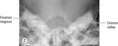
OF 30°
• Foramen magnum is demonstrated
• Dorsum sellae is seen in the centre of the foramen magnum
• Sharp image showing dorsum sellae in contrast with the less dense foramen magnum
| Common errors – OF 20° | Possible reasons |
| Petrous ridge seen above the level of the inferior orbital margins | Inadequate angle selected or OMBL is incorrectly positioned (chin down too far) |
| Petrous ridge seen below the level of the inferior orbital margins | Angle selected is too great or OMBL is incorrectly positioned (chin not far enough down) |
| Common errors – OF 30° | Possible reasons |
| Foramen magnum appears short or is not evident. Dorsum sellae may be visible above the portion of the foramen magnum that is seen | Angle selected is inadequate or OMBL is positioned incorrectly (chin not far enough down) |
| Large foramen magnum seen but curve of the posterior arch of C1 is seen in its lower third, rather than the anvil shape of dorsum sellae | Angle selected is too great or OMBL is not positioned correctly (chin too far down) |
Mastoids
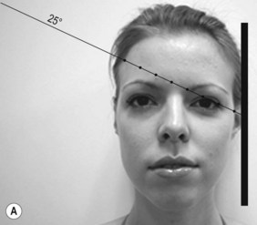
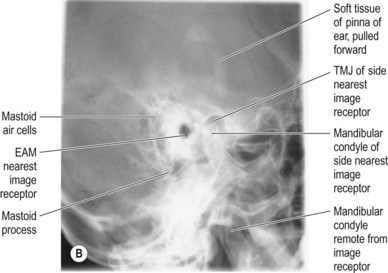
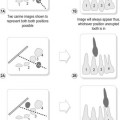
Lateral oblique mastoids (Fig. 20.3A,B)
IR is vertical; an antiscatter grid is employed
Positioning
• The patient is initially seated, facing the IR
• The trunk is brought as close as possible to the receptor unit and the patient is asked to sit with their spine as erect as possible. This helps the patient turn their head more easily into the required lateral position
• The head is turned through 90° to bring the mastoid on the side under examination over the IR. The location of this bone can be detected by palpating the mastoid process, which lies inferiorly and posteriorly to the EAM
• The pinna of the ear on the side nearest the IR is then gently pulled forward and the head rests against the IR to keep the pinna forward. This clears the image of the pinna from the area of interest
Related posts:

Stay updated, free articles. Join our Telegram channel

Full access? Get Clinical Tree


