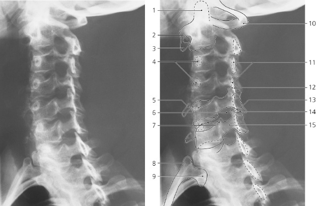
Cervical spine, a-p X-ray
- Foramen transversarium of C III
- Spinous process of C III
- Pedicle of vertebral arch
- Foramen transversarium of C IV
- Superior articular process of C V
- Inferior articular process of C V
- Anterior tubercle of C VI
- Transverse process of C VII
- Pedicle of C VII
- Transverse process of Th I
- Tubercle of first rib
- Head of first rib
- Body of vertebra C V
- Uncus (lip) of C V
- Lamina of thyroid cartilage (calcified)
- Uncovertebral joint (Luschka)
- Spinous process of C VI
- Intervertebral disc C VI – C VII
- Lamina of vertebral arch C VII
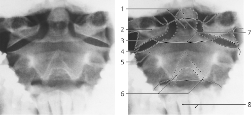
Atlas and axis, a-p X-ray, through open mouth
- Dens axis
- Lateral mass of atlas
- Inferior articular facet of atlas
- Lateral atlanto-axial joint
- Superior articular process of axis
- Spinous process of axis (bifid)
- Anterior and posterior arch of atlas
- Lower incisor teeth
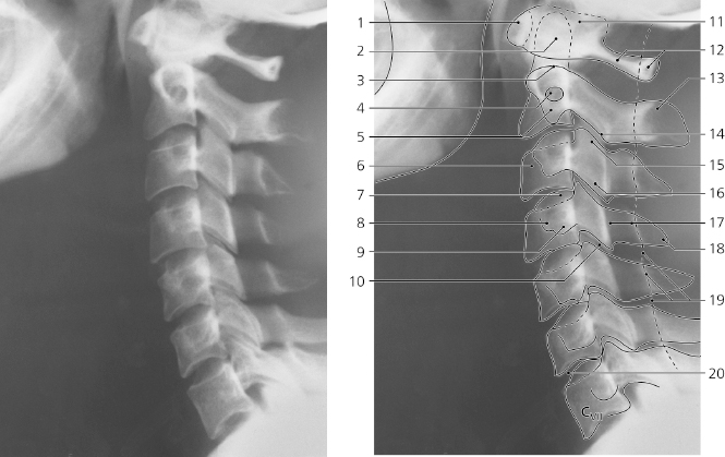
Cervical spine, lateral X-ray
- Anterior arch of atlas
- Dens axis
- Superior articular facet of axis
- Foramen transversarium of axis
- Transverse process of axis
- Body of C III
- Uncus of C IV
- Anterior tubercle of transverse process
- Posterior tubercle of transverse process
- Zygapophysial (facet) joint C IV – C V
- Lateral mass of atlas
- Posterior arch of atlas
- Spinous process of axis
- Inferior articular process of axis
- Superior articular process of C III
- Inferior articular process of C III
- Lamina of vertebral arch C IV
- Spinous process of C IV
- Posterior wall of vertebral canal
- Intervertebral disc C VI – C VII
Cervical spine, oblique X-ray
Only gold members can continue reading.
Log In or
Register to continue

Stay updated, free articles. Join our Telegram channel