Traumatic injuries to the cervical spine comprise approximately 65% of all spinal injuries.
The incidence of cervical spine injury (CSI) after blunt trauma is 2% to 6%.
Cervical spine immobilization is an important initial step in preventing neurologic injury after trauma. The immobilization may be “cleared” by a combination of physical examination findings and radiographic studies.
Computed tomography (CT) has approximately 100% sensitivity for detection of fractures compared with 65% for radiographs, and therefore should be the first imaging modality in patients with suspected injury (Level I evidence).
NEXUS (National Emergency X-Radiography Utilization Study) criteria and CCR (Canadian C-Spine Rule) are validated clinical decision tools to safely rule out CSI without the need to obtain radiographs in alert and stable trauma patients. Both have sensitivity of 90% to 100% for ruling out CSI (Table 2-1).
ACR Appropriateness Criteria rating if imaging indicated by NEXUS criteria:
CT cervical spine without intravenous (IV) contrast—9
Radiographs—6
Magnetic resonance imaging (MRI) without IV contrast for myelopathy—9, and treatment planning for unstable spine—8
Computed tomography angiography head and neck if clinical or imaging findings suggest arterial injury—9
Understanding the mechanism of injury is helpful for evaluating images and determining prognosis (Table 2-2).
Stability of injuries is important to assess. This can be usually accomplished on the lateral view or reformatted CT images, although all images should be assessed.
Indicators of instability include
More than one column involved (Fig. 2-7)
Subluxation greater than 3 mm
Increased interspinous distance
Facet joint widening
Narrowed disc or widened space
Vertebral compression greater than 25%
Not all radiographically documented cervical spine injuries are clinically significant (Table 2-3)
Table 2-1 | ||
|---|---|---|
|

Table 2-2 CERVICAL SPINE TRAUMA: MECHANISMS OF INJURY | ||||||||||||||||||||||||||||
|---|---|---|---|---|---|---|---|---|---|---|---|---|---|---|---|---|---|---|---|---|---|---|---|---|---|---|---|---|
|
Table 2-3 | ||
|---|---|---|
|
 FIGURE 2-1. Disruptive hyperflexion injuries. (A) Mechanism (blow to the occipital region) that causes more posterior soft tissue injury compared with anterior compression. (B) Lateral radiograph shows widened interspinous distance (double arrow), subluxation of the facets (posterior arrowhead) and slight subluxation, and disc space widening (anterior arrowhead). |
 FIGURE 2-2. Compressive hyperflexion injuries. (A) Mechanism of injury with force transmitted to the anterior vertebral body. (B) Lateral radiograph demonstrates compression of C7 and T1 (arrow). |
 FIGURE 2-3. Disruptive hyperextension injuries. (A) Disruptive hyperextension injury with anterior distraction. The cord may be compressed. (B) Lateral radiograph shows anterior disc space widening and vertebral chip fracture (arrow). |
 FIGURE 2-4. Compressive hyperextension injuries. (A) Mechanism resulting in posterior compression and less anterior distraction. (B) Lateral radiograph demonstrates a vertical posterior arch fracture (arrow). |
 FIGURE 2-5. Flexion-rotation injuries. (A) Mechanism of injury. (B) Lateral radiograph shows a unilateral locked facet with subluxation and “bow-tie” configuration (lines) of the facets. |
 FIGURE 2-6. Vertical compression injuries. (A) Mechanism of injury. (B, C) Axial computed tomography (CT) images of a burst fracture. |
 FIGURE 2-6. (continued) |
 FIGURE 2-7. Instability. (A) Lateral radiograph demonstrates the anterior (anterior longitudinal ligament, body, and disc), middle (posterior body and disc and posterior longitudinal ligament), and posterior (facet joints and posterior ligaments) columns. When two columns are involved, the injury should be considered unstable. (B) Lateral view shows multiple column involvement with widened interspinous distance (double arrow), subluxation (black lines), and anterior disc space narrowing (arrowhead) caused by a disruptive hyperflexion injury. |
Atlanto-occipital dislocations are usually fatal; therefore, imaging is rarely performed.
The injury results from high-velocity shearing forces dislocating the head from C1.
In children, the condyles are less well developed, making patients more susceptible to injury.
Occur due to widespread ligamentous disruption between the occiput and upper cervical spine. Often without bony fractures and hence more ready missed by an inexperienced observer.
If the patient survives the injury, treatment is occipital-C2 fusion.
 FIGURE 2-8. Atlanto-occipital dislocation. (A) Lateral radiograph shows a huge prevertebral hematoma (arrows) with anterior dislocation of the occipital condyles (arrowhead). (B) Sagittal computed tomography (CT) reconstruction in another patient following high-speed motor vehicle accident shows clear separation of the occipital condyles from the C1 with huge prevertebral and epidural hematomas and stretching to the cervicomedullary junction. Both patients did not survive. |
Fractures of the atlas (C1) account for 25% of craniocervical injuries, 3% to 13% of cervical spine injuries, and 1% to 3% of all spinal injuries.
Biomechanism: Distraction of C1 lateral masses and failure of the C1 ring at its weakest points adjacent to the lateral masses due to axial loading of occiput.
Isolated posterior arch followed by anterior arch fractures are most common and are managed conservatively with excellent union rates.
Isolated burst fractures (including classic Jefferson fracture) are second most common and seldom cause neurologic injury because the anatomy and mechanism of injury force the fragments away from the spinal canal.
Injuries to transverse ligament are common and can occur without bony injury. Unless the occipital condyles are fractured, other ligaments are spared.
Modified Jefferson Classification:
Type I: Posterior arch alone, most common (67%), bilateral, nondisplaced, and stable
Type II: Anterior arch
Type III: Bilateral posterior arch with unilateral or bilateral anterior arch; includes classic Jefferson burst fracture defined by bilateral fractures of both anterior and posterior arches; usually high-velocity loading mechanism
Type IV: Lateral mass; usually medial portion of lateral mass and unilateral; low-velocity loading mechanism
Type V: Transverse fractures of anterior arch, usually horizontal and in elderly
Forty percent to forty-four percent atlas fractures can occur in combination with axis (C2) or subaxial cervical spine fractures. C2 fractures are usually Type II odontoid or isthmus (hangman’s fractures).
Treatment depends upon integrity of the transverse ligament and the presence or absence of other cervical spine injuries.
Isolated atlas fractures with intact transverse ligament or associated with avulsion of bony medial tubercle have a good union rate with external immobilization (hard collar vs. halo).
Isolated atlas fractures with disruption of mid-portion or nonbony avulsion from medial tubercle cannot heal and are treated with surgical fixation.
Imaging:
Radiographs include open-mouth odontoid, lateral, and flexion-extension views. CT preferred for complete evaluation. MRI highly sensitive for evaluation of transverse ligament rupture.
Displacement of C1 lateral mass over C2 facets suggests burst fracture of atlas.
C1 lateral mass overhang upon C2 facets combined on both sides >7 mm (rule of Spence) is suggestive of ruptured transverse ligament. However, rule of Spence can miss transverse ligament disruption from avulsion of medial tubercle.
The amount of C1 lateral mass overhang is important for surgical planning and fixation approach (transarticular vs. lateral mass).
Atlantodental interspace >3 mm in adults and >5 mm in children highly suggestive of ruptured transverse ligament.
Retropharyngeal soft tissue swelling at C1-C3 suggestive of anterior arch fracture.
 FIGURE 2-9. Jefferson fracture. (A) Mechanism of injury for Jefferson fractures. (B) Anteroposterior (AP) open-mouth odontoid view shows displacement of the lateral masses outward (arrows). The extent of injury is best appreciated on the axial (C) and coronal (D) computed tomography (CT) images. |
 FIGURE 2-10. Hyperextension injury C1. Lateral radiograph of the upper cervical spine demonstrates an avulsion fracture of the anterior arch of C1 (arrow). |
Traumatic dislocations of the atlantoaxial axis are rare.
The transverse ligament assists in maintaining the normal C1-C2 relationship. Subluxations may occur with conditions that affect the normal osseous or ligamentous integrity (rheumatoid arthritis, retropharyngeal infection, congenital abnormalities, and trauma).
Traumatic subluxation/dislocation may be anterior, posterior, or rotary in nature.
Normally, the space between the odontoid and lower anterior arch of C1 is 2 mm in adults and 4 mm in children.
Imaging can be accomplished with lateral and open-mouth odontoid views. However, thin-section CT with reformatting or three-dimensional reconstruction is most accurate for detection and classification.
 FIGURE 2-11. (A) Lateral radiograph of the upper cervical spine demonstrates anterior subluxation of C1 on C2. Odontoid-C1 and posterior arch relationships (dotted lines). (B) Axial computed tomography (CT) image demonstrates an anterior arch fracture (arrow) and avulsion (arrowhead) of the transverse ligament. |
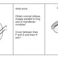
 FIGURE 2-12. Rotary subluxation/fixation. Skeletal specimens with metal markers on the C2 facet demonstrate normal (A) and rotary fixation (B) of C1 on C2. Axial computed tomography (CT) (C) and three-dimensional surface rendered reconstructions, caudocranial (D) and craniocaudal (E) views show rotary fixation. The rotation of C1 (solid line) in relation to C2 (broken line) and (O) odontoid process in (C). |
 FIGURE 2-12. (continued) |
Fractures of C2 are common, accounting for 15% to 27% of cervical spine injuries (Table 2-4). Up to 25% have associated lower cervical fractures. Odontoid fractures account for 75% of cervical spine injuries in children.
Ossification of the dens completes by 12 years of age; this must be considered in pediatric trauma cases.
Fractures of the odontoid and neural arch (hangman’s fracture) account for 80% of C2 fractures.
Odontoid fractures are classified by location (Table 2-5). Fractures of the base of the odontoid (Type II) have a high incidence of nonunion (54% to 67%) and instability.
Hangman’s fractures (neural arch) are hyperextension injuries.
Posterior C1-C2 fixation methods have a high fusion rate.
Anterior odontoid screw placement is contraindicated in cases of chronic fracture, disruption of transverse ligament, or angulated displacement of dens.
Table 2-4 AXIS (C2) FRACTURES | ||||||||||
|---|---|---|---|---|---|---|---|---|---|---|
|
Table 2-5 ODONTOID FRACTURE CLASSIFICATION | ||||||||||||
|---|---|---|---|---|---|---|---|---|---|---|---|---|
|
 FIGURE 2-13. (A) Odontoid fracture classification: Type I—odontoid tip; Type II—odontoid base, above accessory ligament and vascular supply (arrows); and Type III—extend into body below vascular supply. Sagittal and coronal computed tomography (CT) images; (B, C) Type I (chronic) fracture; (D, E) Type II fracture with posterior subluxation of C1 and displacement of the odontoid process; and (F, G) Type III fracture. |
 FIGURE 2-14. Hangman’s fracture with anterior displacement of C2 on C3 on lateral radiograph (A) and sagittal computed tomography (CT) image (B). Fracture of bilateral C2 pars on axial CT (C). |
 FIGURE 2-15. Anterior inferior body fracture of C2 (arrow). |
Vertebral arch fractures are most frequently the result of hyperextension or lateral flexion injuries. Common sites and incidences in our experience are as follows:
Lamina
28%
Pedicle
25%
Spinous process
22%
Pillar
16%
Facets
9%
Transverse process
2%
Laminar fractures are commonly associated with spinous process fractures. The majority occur from C5-C7.
Pedicle fractures may occur at C2 (hangman’s) or in the lower cervical spine. If nondisplaced, they may be easily overlooked on routine radiographs.
Spinous process fractures are most common from C5-T1. Injury may be the result of direct trauma, hyperextension, or hyperflexion.
Pillar fractures without displacement may be overlooked on radiographs. They are most common at C6.
Facet fractures are the result of flexion-rotation or hyperflexion injuries.
Transverse process fractures occur with direct trauma or lateral flexion, and are managed conservatively.
In general, isolated nondisplaced posterior element fractures have a high spontaneous union rate and are managed conservatively with bracing unless there is neurologic compromise.
Displaced fractures of the facet or pedicle are treated with surgical fixation; however, conservative treatment may be attempted.
Transverse process fractures are stable; however, it is important to assess for potential vertebral artery injury secondary to transverse foramen compromise (40% incidence).
 FIGURE 2-16. Computed tomography (CT) image of a remote fracture of the left transverse process through the foramen transversarium (arrows). |
 FIGURE 2-17. Sagittal computed tomography (CT) image of a C5 facet fracture (arrow). |
 FIGURE 2-18. Spinous process fracture at C7 (Clay shoveler’s fracture). (A) Anteroposterior (AP) view shows a double spinous process (arrows). This is a helpful sign because the lower cervical spine is often difficult to visualize on the lateral view. (B) Lateral view shows the C7 spinous process fracture clearly in this case. |
More than 31% of cervical spine injuries involve the vertebral body. Seventy-five percent of fractures occur from C5-C7.
Flexion teardop fractures (avulsion injuries of inferior vertebral body) may have a benign appearance on plain radiographs; however, they have a high rate of spinal cord injury and are unstable injuries.
Burst fractures usually occur in the lower cervical spine and have a high incidence of neurologic injury; however, they may be managed conservatively with bracing if there is no ligamentous disruption.
Ligamentous disruption can be hidden by spontaneous reduction in plain radiographs taken in the supine position.
Table 2-6 summarizes vertebral body fractures.
Table 2-6 VERTEBRAL BODY FRACTURES | |||||||||||||||||||||||
|---|---|---|---|---|---|---|---|---|---|---|---|---|---|---|---|---|---|---|---|---|---|---|---|
| |||||||||||||||||||||||
 FIGURE 2-19. Flexion-compression injury with compression of C7 and an anterior chip fracture (open arrow). There is widening of the interspinous distance (arrow) indicating posterior ligament injury and instability. |
 FIGURE 2-20. Teardrop fractures. (A) Hyperextension teardrop of C4. Note the posterior arch fractures of C2 and C3 (arrows) indicating the mechanism of injury. (B) Hyperflexion teardrop fracture of C5 with compromise of the spinal canal (dotted lines) and posterior ligament tears (double arrow)—finding best seen on sagittal computed tomography (CT) image (C). Axial CT (D) shows the sagittal fracture commonly seen with flexion teardrop fracture. |
 FIGURE 2-21. Axial (A) and sagittal (B) computed tomography (CT) images show chronic C5 vertebral body burst fracture with retropulsion which mildly narrows the spinal canal. Also noted are chronic compression deformities of C6 and C7 vertebral bodies and a nonunited Type II odontoid fracture. |
Most significant ligament injuries involve the posterior column. Seventy-three percent occur at the C5-C7 levels. Anterior ligament injuries are most common at C2-C4 and C6-C7. Ligament injuries may occur alone or with fractures.
The spectrum of injuries may be subtle, without obvious abnormality and normal bony alignment, or injuries may be grossly obvious, such as bilateral locked facets.
Unilateral facet locking or perching may present with subtle radiographic findings. This flexion-rotation injury accounts for 12% of cervical injuries. Reformatted CT imaging is often required for detection and complete evaluation of these injuries.
Flexion-distraction injuries may present with acute spinal cord injury, radiculopathy, or both.
Magnetic resonance (MR) should be performed in stable patients who have suspected ligamentous injury and inconclusive findings on CT.
Ligamentous injury (anterior longitudinal ligament [ALL] and posterior longitudinal ligament [PLL]) is an unstable injury, and open reduction/internal fixation is indicated.
 FIGURE 2-22. Flexion-distraction injury with posterior ligament tear at C5-C6. Flexion (A) and extension (B) views show subluxation and widening of the interspinous distance and facet joints with flexion that reduces with extension. Treatment—posterior fusion. |
 FIGURE 2-23. Unilateral locked facet. (A) Anteroposterior (AP) radiograph shows disc space asymmetry (arrow) at C4-C5 and rotation of the spinous process. (B) Lateral view shows subluxation, and the C4 facets form the “bow-tie” sign. (C) Oblique view shows the facet overlap (arrow). |
 FIGURE 2-24. Sagittal computed tomography (CT) images of locked (A) and perched (B) facets (arrow). |
 FIGURE 2-25. Lateral radiograph (A) demonstrates bilateral locked facets at C5-C6 with a chip fracture (arrow) of C6. Subluxation with bilateral locked facets must be equal or greater than 50%. Sagittal T2 magnetic resonance imaging (MRI) (B) in another patient with bilateral facet dislocation demonstrates spinal cord impingement, ligamentum flavum, and interspinous ligament injury (black arrows) with resultant fanning of the C6 and C7 spinous processes. Quadriplegia occurs in 72% of patients with this injury. |
Because of differences in range of motion, transition in the facet joints, and other anatomic factors, the thoracolumbar junction is most susceptible to injury (66% of injuries occur between T12 and L2).
Mechanism of injury
Hyperflexion (most common)
Flexion-compression
Flexion-distraction
Flexion-rotation
Lateral flexion
Vertical compression
Hyperextension
Shearing/rotation forces
Complete evaluation of bone and soft tissue structures is essential to assess stability.
Thoracolumbar spine injuries are classified on the basis of radiographic and CT features.
Instability: Three-column approach of Denis is most useful. If two columns (Fig. 2-26) are involved, the injury is unstable.
Image evaluation: Routine radiographs remain the primary screening tool for thoracolumbar fractures. However, CT is almost 100% sensitive for fracture detection compared with 32% for radiographs. CT is also useful to fully evaluate fragment position and spinal canal compromise. MRI is useful in selected cases to evaluate the cord, nerve roots, and soft tissue injuries. Unsuspected posterior ligament injuries are evident on MRI, when not seen on radiographs or CT.Related posts:

Stay updated, free articles. Join our Telegram channel

Full access? Get Clinical Tree