Benign Tumors and Tumor-like Lesions II: Lesions of Cartilaginous Origin
Benign Chondroblastic Lesions
Diagnosis of a bone lesion as originating from cartilage is usually a simple task for the radiologist. The lesion’s radiolucent matrix, scalloped margins, and annular, comma-shaped, or punctate calcifications usually suffice to establish its chondrogenic nature. However, whether a cartilage tumor is benign or malignant is sometimes extremely difficult for the radiologist to determine.
Enchondroma (Chondroma)
Enchondroma is the second most common benign tumor of bone, constituting approximately 10% of all benign bone tumors and representing the most common tumor of the short tubular bones of the hand. This benign lesion is characterized by the formation of mature hyaline cartilage. When it is located centrally in the bone, it is termed an enchondroma (Fig. 18.1); if it is extracortical (periosteal) in location, it is called a chondroma (periosteal or juxtacortical) (see Figs. 18.10 and 18.11). Although occurring throughout life, enchondromas are usually seen in patients in their second through fourth decades. There is no sex predilection. The short tubular bones of the hand (phalanges and metacarpals) are the most frequent sites of occurrence (Fig. 18.2), although the lesions are also encountered in the long tubular bones (Fig. 18.3). According to recent investigations, enchondromas result from the continued growth of residual benign cartilaginous rests that are displaced from the growth plate. They are often asymptomatic; a pathologic fracture through the tumor (Figs. 18.4 and 18.5) often calls attention to the lesion.
Enchondroma protuberans is a rare variant. It is a lesion that arises in the intramedullary cavity of a long bone and forms a prominent exophytic mass on the cortical surface. This lesion must be distinguished from an osteochondroma or central chondrosarcoma that penetrates the cortex and forms a juxtacortical mass.
In most instances, radiography suffices to demonstrate the lesion. In the short bones, the lesion is often entirely radiolucent (Fig. 18.6), whereas in the long bones it may display visible calcifications. If the calcifications are extensive, enchondromas are called “calcifying” (Fig. 18.7). The lesions can also be recognized by shallow scalloping of the inner (endosteal) cortical margins because the cartilage in general grows in a lobular pattern (see Fig. 18.1).
Computed tomography (CT) and magnetic resonance imaging (MRI) may further delineate the tumor and more precisely localize it in the bone. On spin-echo T1-weighted MR images, enchondromas demonstrate intermediate to low signal intensity, whereas on T2-weighted images they exhibit high signal intensity. The calcifications within the tumor will image as low-signal-intensity structures (Figs. 18.8 and 18.9). It must be stressed, however, that most of the time neither CT nor MRI is suitable for establishing the precise nature of a cartilaginous lesion, nor can CT or MRI distinguish benign from malignant lesions. Despite the use of various criteria, the application of MRI to the tissue diagnosis of cartilaginous lesions has not brought satisfactory results, although preliminary results of recent trials with fast contrast-enhanced MR imaging showed that this technique might assist in differentiation between the benign and malignant cartilaginous tumors.
Skeletal scintigraphy usually reveals mild to moderate increased uptake of the tracer in uncomplicated enchondromas, whereas the presence of a pathologic fracture or malignant transformation is revealed by marked scintigraphic activity.
Intracortical chondroma is a very rare variant of conventional enchondroma. The lesion is located in cortical bone and is surrounded by sclerosis of the medullary bone and periosteal reaction. Some of these lesions may actually represent periosteal chondroma with an atypical radiographic appearance, as reported by Abdelwahab and associates. Intracortical chondroma can occasionally simulate an osteoid osteoma.
Periosteal chondroma is a slow-growing, benign cartilaginous lesion that arises on the surface of a bone in or beneath the periosteum. It occurs in children as well as adults, with no sex predilection. There is usually a history of pain and tenderness, often accompanied by swelling at the site of the lesion, which is most commonly located in the proximal humerus. As the tumor enlarges, it is seen radiographically eroding the cortex in a saucer-like fashion, producing a solid buttress of periosteal new bone (Fig. 18.10). The lesion has a sharp sclerotic inner margin demarcating it from the buttress of periosteal new bone. Scattered calcifications are often seen within the lesion (Fig. 18.11).
CT may show to better advantage the scalloped cortex and matrix calcification (Fig. 18.12). It also may demonstrate the separation of a lesion from the medullary cavity, an important feature
in differentiation from osteochondroma. MRI findings correspond to radiographic findings, depicting the cartilaginous soft-tissue component. If periosteal chondroma affects the medullary canal, MRI may be useful in depicting the extent of involvement (Fig. 18.13). Fat suppression or enhanced gradient-echo sequences may improve tumor-marrow contrast. The potential pitfall of MRI is marrow edema mimicking tumor invasion or vice versa. Unlike enchondroma and osteochondroma, periosteal chondroma may continue to grow after skeletal maturation. Some lesions may attain a large size (up to 6 cm) and may resemble osteochondromas (Figs. 18.14 and 18.15). Some lesions may mimic an aneurysmal bone cyst. Very rarely the lesion may encase itself intracortically, thus mimicking other intracortical
lesions (such as intracortical angioma, intracortical fibrous dysplasia, or intracortical bone abscess).
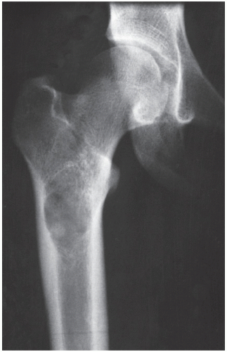 FIGURE 18.1 Enchondroma. A radiolucent lesion in the medullary portion of the proximal femur of a 22-year-old man is seen eroding the inner aspect of the lateral cortex. Note scalloped borders and matrix calcification. |
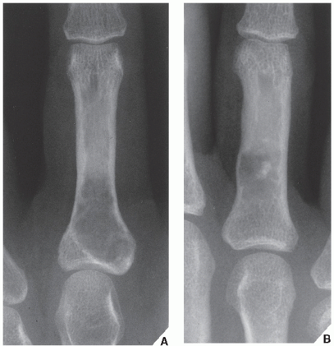 FIGURE 18.2 Enchondroma. (A) A radiolucent lesion in the proximal phalanx of the middle finger of a 40-year-old woman, and (B) a similar lesion with central calcification in the proximal phalanx of the ring finger of a 42-year-old man are typical examples of enchondroma in the short tubular bones. |
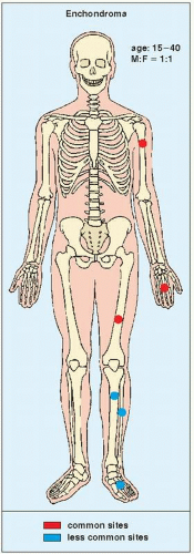 FIGURE 18.3 Skeletal sites of predilection, peak age range, and male-to-female ratio in enchondroma. |
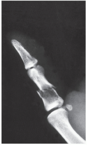 FIGURE 18.4 Enchondroma. Radiograph of a 31-year-old man who had injured his left thumb reveals a pathologic fracture through an otherwise asymptomatic lesion. |
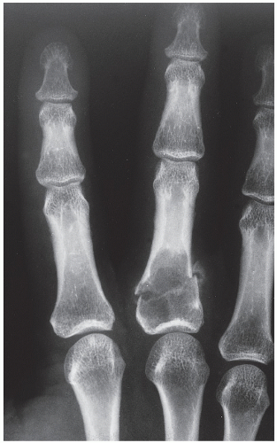 FIGURE 18.5 Enchondroma. Pathologic fracture through a large enchondroma is present in the proximal phalanx of the middle finger. |
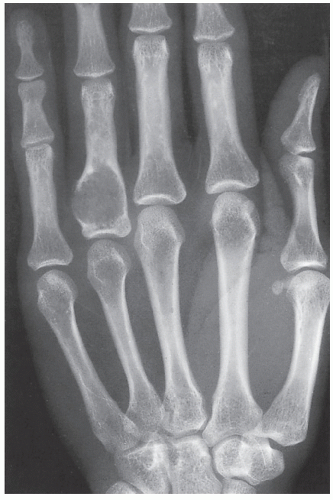 FIGURE 18.6 Enchondroma. A typical, purely radiolucent lesion at the base of the proximal phalanx of the ring finger of a 37-year-old woman represents an enchondroma. Note the marked attenuation of the ulnar side of the cortex. |
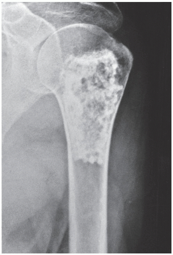 FIGURE 18.7 Calcifying enchondroma. In this heavily calcified enchondroma of the proximal humerus of a 58-year-old woman, note the lobular appearance of the lesion and the minimal degree of scalloping of the lateral endocortex. |
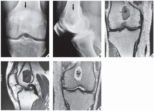 FIGURE 18.8 MRI of enchondroma. Anteroposterior (A) and lateral (B) radiographs of the left knee of a 61-year-old man demonstrate only a few calcifications in the distal femur (arrows). The extent of the lesion cannot be determined. Coronal (C) and sagittal (D) T1-weighted MR images show a well-circumscribed, lobulated lesion displaying intermediate signal intensity. The darker area in the center represents calcifications. Coronal T2-weighted image (E) shows the lesion displaying a mixed-intensity signal: the brighter areas represent cartilaginous tumor and the darker areas calcifications. |
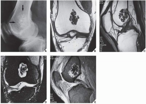 FIGURE 18.9 MRI of enchondroma. (A) Lateral radiograph of the knee shows chondroid calcifications in the distal femur (arrows). Coronal (B) and sagittal (C) spin-echo T1-weighted MR images show the lesion being predominantly of low signal intensity. Coronal (D) inversion recovery T2-weighted with fat saturation and sagittal (E) fast-spin echo T2-weighted images demonstrate the full extent of enchondroma. Calcifications exhibit low signal intensity. |
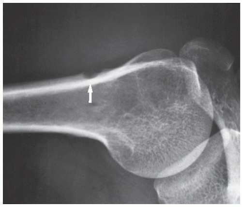 FIGURE 18.10 Periosteal chondroma. A radiolucent lesion (arrow) is eroding the external surface of the cortex of the proximal humerus of a 24-year-old man. |
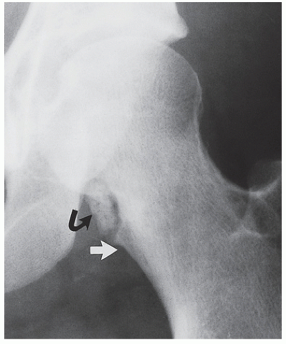 FIGURE 18.11 Periosteal chondroma. A periosteal chondroma at the medial aspect of the neck of the left femur eroded the cortex in a saucer-like fashion. The characteristic buttress of a periosteal reaction is seen at the inferior border of the lesion (arrow). Note also cluster of calcification in the soft tissue (curved arrow). |
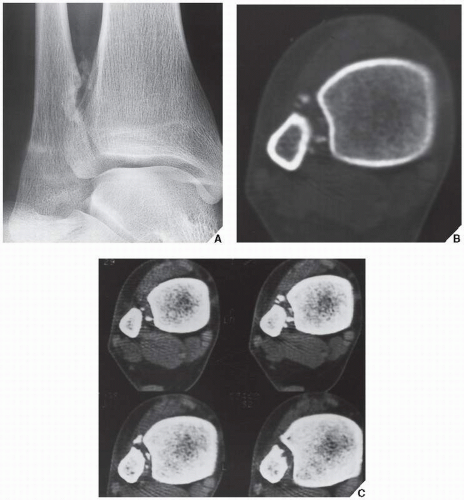 FIGURE 18.12 CT of periosteal chondroma. (A) An oblique radiograph of the right ankle shows a lesion containing calcifications eroding the medial cortex of the distal fibula. CT using a bone window (B) and a soft-tissue window (C) better demonstrates the extent of the lesion and the distribution of the calcifications. |
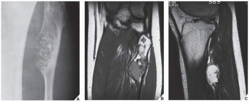 FIGURE 18.13 MRI of periosteal chondroma. (A) A large periosteal chondroma eroded the cortex of the proximal fibula and extended into the medullary cavity. Coronal (B) proton-density (SE; TR 2000/TE 19 msec) and sagittal (C) T2-weighted (SE; TR 2000/TE 70 msec) MRI show the lesion’s extension into the bone marrow. |
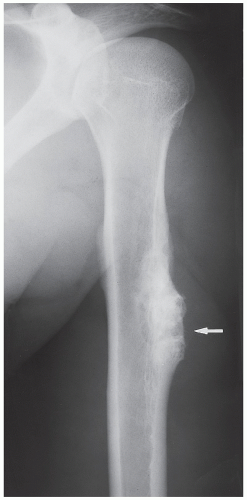 FIGURE 18.14 Periosteal chondroma. A large periosteal chondroma (arrow) mimics an osteochondroma. Note, however, the periosteal reaction and separation of the tumor from the medullary cavity by a cortex, features that helped in the differentiation from osteochondroma. (Courtesy of Dr. KK Unni, Rochester, Minnesota.) |
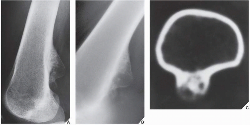 FIGURE 18.15 Periosteal chondroma. (A) Lateral radiograph of the distal femur shows a lesion arising from the posterior cortex that resembles an osteochondroma. (B) Conventional tomography shows calcifications at the base of the lesion and continuity of the posterior cortex of the femur. (C) CT section demonstrates lack of communication between the medullary portion of the femur and the lesion, thus excluding the diagnosis of osteochondroma (A and C from Greenspan et al., 1993, with permission.) |
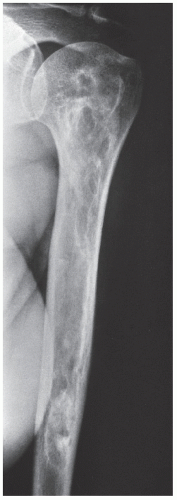 FIGURE 18.16 Bone infarct. In a medullary bone infarct, seen here in the proximal humerus of a 36-year-old man with sickle-cell disease, there is no endosteal scalloping of the cortex, and the calcified area is surrounded by a thin, dense sclerotic rim, the hallmark of a bone infarct. |
Histologically, enchondroma consists of lobules of hyaline cartilage of varying cellularity and is recognized by the features of its intracellular matrix, which has a uniformly translucent appearance and contains relatively little collagen. The tissue is sparsely cellular, and the cells contain small and darkly staining nuclei. The tumor cells are located in rounded spaces known as lacunae. On histologic examination of periosteal chondroma, the findings are identical to those of enchondroma, although the lesion sometimes exhibits higher cellularity, occasionally with atypical cells.
Differential Diagnosis
The main differential diagnosis of enchondroma, particularly in lesions of the long bones, is a medullary bone infarct (Fig. 18.16). At times, the two lesions may be difficult to distinguish from one another, particularly if the enchondroma is small, because both lesions present with similar calcifications. The radiographic features helpful in the differential diagnosis are the lobulation of the inner cortical margins in enchondroma, the annular, punctate, and comma-shaped calcifications in the matrix, and the lack of sclerotic rim that is usually seen in bone infarcts (Fig. 18.17).
The most difficult task for the radiologist is to distinguish a large solitary enchondroma from a slowly growing low-grade chondrosarcoma. One of the most significant findings pointing to a chondrosarcoma in the early stage of development is localized thickening of the cortex (Fig. 18.18). The size of the lesion should also be taken into consideration. Lesions longer than 4 cm (or, according to some investigators, longer than 7 cm) are suggestive of malignancy. In more advanced tumors, destruction of the cortex and the presence of a soft-tissue mass are the hallmarks of malignancy.
Complications
The single most important complication of enchondroma, aside from pathologic fracture (see Fig. 18.4), is its malignant transformation to chondrosarcoma. With solitary enchondromas, this occurs almost
exclusively in a long or flat bone and almost never in a short tubular bone. The radiographic signs of the transformation are thickening of the cortex, destruction of the cortex, and a soft-tissue mass. The development of pain in the absence of fracture at the site of the lesion is an important clinical sign.
exclusively in a long or flat bone and almost never in a short tubular bone. The radiographic signs of the transformation are thickening of the cortex, destruction of the cortex, and a soft-tissue mass. The development of pain in the absence of fracture at the site of the lesion is an important clinical sign.
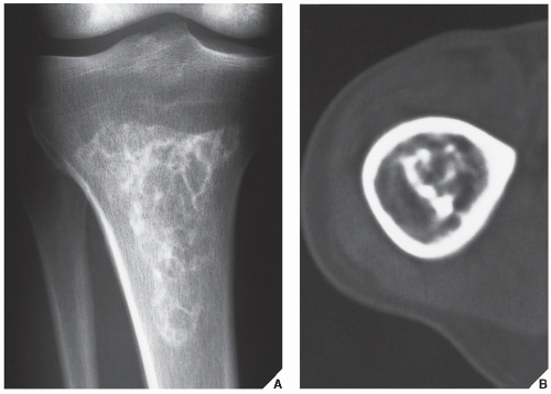 FIGURE 18.17 Bone infarct. (A) Conventional radiograph of the proximal tibia shows the typical coarse calcifications of medullary bone infarct. Note the sharply defined peripheral margin separating necrotic from viable bone, and the lack of characteristics for chondroid tumor annular and comma-shaped calcifications. (B) In another patient with a bone infarct in the distal femur, a CT section reveals central coarse calcifications and no endosteal scalloping of the cortex. |
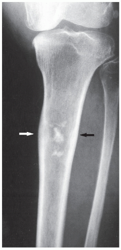 FIGURE 18.18 Low-grade chondrosarcoma. A 48-year-old woman presented with pain in the upper leg. A radiograph shows a radiolucent lesion in the proximal tibia with a wide zone of transition and central calcifications. Note focal thickening of the cortex (arrows), an important feature that distinguishes chondrosarcoma from similarly appearing enchondroma. |
Treatment
Curettage of the lesion with the application of bone graft is the most common course of treatment.
Enchondromatosis (Ollier Disease)
Enchondromatosis is a condition marked by multiple enchondromas, generally in the region of the metaphysis and diaphysis (Fig. 18.19). If the skeleton is extensively affected, with predominantly unilateral distribution, the term Ollier disease is applied. The clinical manifestations of multiple enchondromas, such as knobby swellings of the digits or gross disparity in the length of the forearms or legs, are frequently recognized in childhood and adolescence; the disease has a strong preference for one side of the body. The disorder has no hereditary or familial tendency. Some investigators claim that it is not a neoplastic lesion but rather a developmental bone dysplasia.
The pathogenesis of Ollier disease is unknown. There are two hypotheses for the mechanism of enchondroma formation—one attributes formation to ectopic nests of chondroblasts and the other to the failure of chondrocytes and the growth plate to mature.
Conventional radiography is usually sufficient to demonstrate the typical features of enchondromatosis. Characteristically, interference of the lesion with the growth plate causes foreshortening of the limbs. Deformity of the bones is marked by radiolucent masses of cartilage, often in the hand and foot, containing foci of calcification (Fig. 18.20). Enchondromas in this location may be intracortical and periosteal. They sometimes protrude from the shaft of the short or long tubular bone, thus resembling osteochondromas (Fig. 18.21). Linear columns of cartilage in the form of radiolucent streaks extend from the growth plate to the diaphysis, and a fan-like pattern is common in the iliac bones (Fig. 18.22).
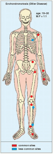 FIGURE 18.19 Skeletal sites of predilection, peak age range, and male-to-female ratio in enchondromatosis (Ollier disease). |
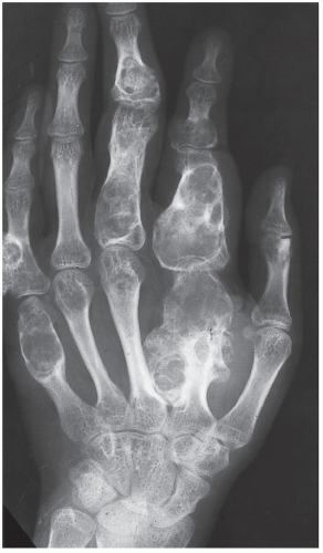 FIGURE 18.20 Ollier disease. Large, lobulated cartilaginous masses markedly deform the bones of the hand in this 20-year-old man. |
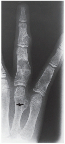 FIGURE 18.21 Enchondromatosis. In this 12-year-old boy, the intracortical lesion in the metaphysis of the fourth metacarpal protrudes from the bone (arrow), thus resembling an osteochondroma. |
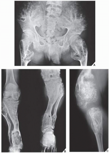 FIGURE 18.22 Ollier disease. The classic features of Ollier disease in a 17-year-old boy are exhibited in extensive involvement of multiple bones. (A) Anteroposterior radiograph of the pelvis demonstrates crescent-shaped and ring-like calcifications in tongues of cartilage extending from the iliac crests and proximal femora. (B) A radiograph of both legs shows growth stunting and deformities of the tibia and fibula. (C) In another patient, a 6-year-old boy with Ollier disease, note extensive involvement of the tibia and distal femur. (A from Norman A, Greenspan A, 1982, with permission.) |
Histologically, the lesions of enchondromatosis are essentially indistinguishable from those of solitary enchondromas, although on occasion they tend to be more cellular.
Complications
The most frequent and severe complication of Ollier disease is malignant transformation to chondrosarcoma. In contrast to solitary enchondromas, even lesions in the short tubular bones may undergo sarcomatous change (Fig. 18.23). This is particularly true in patients with Maffucci syndrome, a congenital, nonhereditary disorder manifested by enchondromatosis and soft-tissue hemangiomatosis (Fig. 18.24). The hemangiomas are mostly located in the subcutaneous soft tissues. The skeletal lesions in this syndrome show a predilection for involvement of the tubular bones and have the same distribution as those in Ollier disease, with a similarly strong predilection for one side of the body. Maffucci syndrome is recognized radiographically by multiple calcified phleboliths.
Osteochondroma
Also known as osteocartilaginous exostosis, this lesion is characterized by a cartilage-capped bony projection on the external surface of a bone. It is the most common benign bone lesion, constituting approximately 20% to 50% of all benign bone tumors, and is usually diagnosed in patients before their third decade. Osteochondroma, which has its own growth plate, usually stops growing at skeletal maturity. The most common sites of involvement are the metaphyses of the long bones, particularly in the region around the knee and the proximal humerus (Fig. 18.25). Variants of osteochondroma include subungual exostosis, turret exostosis,
traction exostosis, bizarre parosteal osteochondromatous proliferation, florid reactive periostitis, and dysplasia epiphysealis hemimelica (also called intraarticular osteochondroma).
traction exostosis, bizarre parosteal osteochondromatous proliferation, florid reactive periostitis, and dysplasia epiphysealis hemimelica (also called intraarticular osteochondroma).
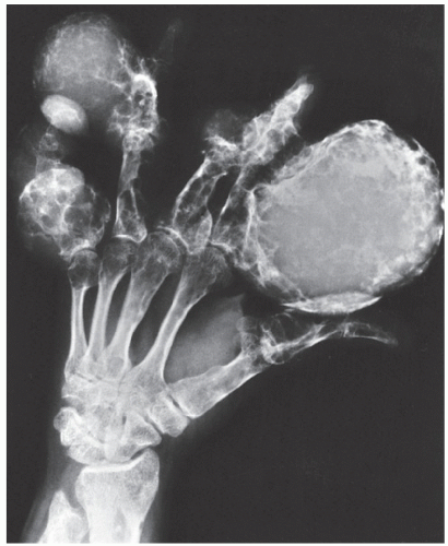 FIGURE 18.23 Chondrosarcoma in Ollier disease. In this case of sarcomatous transformation of enchondroma in the hand in a patient with Ollier disease, note the large, lobulated masses of cartilage in all fingers. The lesion of the middle phalanx of the ring finger shows destruction of the cortex and extension into the soft tissues. |
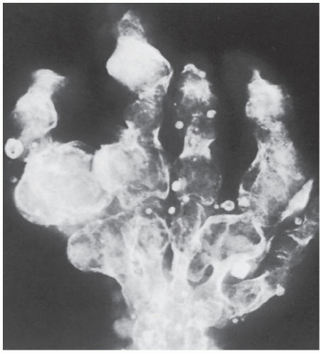 FIGURE 18.24 Maffucci syndrome. Radiograph of the hand reveals typical changes of enchondromatosis, accompanied by calcified phleboliths in soft-tissue hemangiomas. (From Bullough PG, 1992, with permission.) |
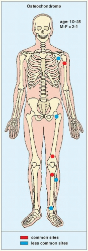 FIGURE 18.25 Skeletal sites of predilection, peak age range, and male-to-female ratio in osteochondroma (osteocartilaginous exostosis). |
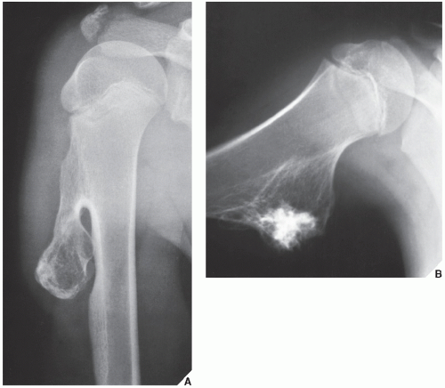 FIGURE 18.26 Osteochondroma. (A) The typical pedunculated type of osteochondroma is seen arising near the proximal growth plate of the right humerus in a 13-year-old boy. (B) In the typical sessile or broad-based variant, seen here arising from the medial cortex of the proximal diaphysis of the right humerus in a 14-year-old boy, the cortex of the host bone merges without interruption with the cortex of the lesion. The cartilaginous cap is not visible on the conventional radiographs, but dense calcifications in the stalk can be seen. |
The radiographic presentation of osteochondroma is characteristic according to whether the lesion is pedunculated, with a slender pedicle usually directed away from the neighboring growth plate (Fig. 18.26A), or sessile, with a broad base attached to the cortex (Fig. 18.26B). The most important characteristic feature of either type of lesion is uninterrupted merging of the cortex of the host bone with the cortex of the osteochondroma; additionally, the medullary portion of the lesion and the medullary cavity of the adjacent bone communicate. CT scanning can establish unequivocally the lack of cortical interruption and the continuity of cancellous portions of the lesion and the host bone (Fig. 18.27). These are important features that distinguish this lesion from the occasionally similar looking bone masses of osteoma, periosteal chondroma, juxtacortical osteosarcoma, soft-tissue osteosarcoma, and juxtacortical myositis ossificans (Fig. 18.28). The other characteristic feature of osteochondroma involves calcifications in the chondro-osseous portion of the stalk of the lesion (see Fig. 18.26) and cartilaginous cap. The thickness of the cartilaginous cap ranges from 1 to 3 mm and rarely exceeds 1 cm. On MRI, the cartilaginous cap shows high signal intensity on T2-weighted and gradient-echo sequences. A narrow band of low signal intensity surrounding the cap represents the overlying perichondrium (Fig. 18.29).
Related posts:


Stay updated, free articles. Join our Telegram channel

Full access? Get Clinical Tree


