Radiologic Evaluation of Skeletal Anomalies


Radiologic Evaluation of Skeletal Anomalies


Classification

The conditions discussed in this part comprise disturbances in skeletal formation, development, growth, maturation, and modeling. Some of these anomalies arise during fetal development, such as congenital absence of a whole or part of a limb, supernumerary digits in a hand or foot, or fused digits, and are obvious at the time the baby is born. Some may begin to develop during fetal life but become apparent later in childhood, such as Hurler syndrome (gargoylism) or osteogenesis imperfecta tarda. Other anomalies, such as certain sclerosing dysplasias, develop after birth because of a genetic predisposition and become manifest later in life.

Congenital anomalies can be classified in various ways, but because of their complexity a full and detailed classification of these disorders is beyond the scope of this chapter. To simplify the variety of classifications, which are constantly changing and expanding, the congenital anomalies may be divided from the pathologic point of view into those involving disturbances of bone formation, bone growth, and bone maturation and modeling (Table 31.1). Anomalies of bone formation include the complete failure of a bone to form and faulty formation of bones, which may manifest in a decreased number of bones (agenesis and aplasia) (Fig. 31.1A,B) or in the number of supernumerary bones (polydactyly) (Fig. 31.1C,D). Anomalies of formation may also be encountered in aberrations involving bone differentiation, which include pseudoarthroses (Fig. 31.2A) and bone fusions (syndactyly and synostosis) (Fig. 31.2B-E). Disturbances in bone growth may lead to aberrations in the size or shape of bones. These may manifest in undergrowth (hypoplasia or atrophy) (Fig. 31.3A-C), overgrowth (hypertrophy or gigantism) (
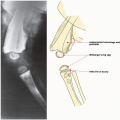
Fig. 31.3D), or deformed growth, such as congenital tibia vara (see Fig. 32.40). Anomalies related to bone growth may also be exhibited in abnormalities affecting the motion in a joint, such as contractures, subluxations, and dislocations (Fig. 31.4). Among the last group of congenital anomalies affecting the skeletal system are those exhibiting aberrations in bone growth, maturation, and modeling, as manifest in the various dysplasias (Fig. 31.5).

A second simple classification system is anatomic and based on the affected region of the body. This system comprises anomalies of the shoulder girdle and upper limb, pelvis and lower limb, spine, and the skeleton in general.


Radiologic Imaging Modalities

Radiologic examination is essential for the accurate diagnosis of many congenital and developmental anomalies, which in some instances (such as osteopoikilosis or osteopathia striata) are totally asymptomatic and only revealed on radiographs obtained for other purposes. It also plays an important part in monitoring the progress of treatment. In many instances the results of therapy, whether conservative or surgical, can be assessed only on the basis of the proper radiologic examination.

The radiologic imaging modalities most frequently used in diagnosing congenital malformations of the bones and joints are the following:


  • Conventional radiography, including standard and special projections


  • Arthrography


  • Myelography


  • Computed tomography (CT)


  • Radionuclide imaging (scintigraphy, bone scan)


  • Ultrasound (US)


  • Magnetic resonance imaging (MRI)

In most instances, the diagnosis can be made on the standard radiographic projections specific for the anatomic site under investigation. As in most other orthopedic conditions, radiographs should be obtained in at least two projections at 90 degrees to one another (Fig. 31.6, see also Fig. 4.1). Supplemental views, however, are sometimes necessary for a full evaluation of an anomaly, particularly those affecting complex structures such as the ankle and foot (Fig. 31.7). Weight-bearing radiographs of the foot should be obtained whenever possible.

Ancillary imaging techniques play an important role in the evaluation of many congenital and developmental conditions. Myelography, for example, is still valuable for detecting anomalies of the spine (Fig. 31.8). In congenital dislocations, particularly in the hip, arthrography remains an essential technique (Fig. 31.9); it is also effective in demonstrating developmental anomalies affecting the articular cartilage and menisci of the knee, as in Blount disease (Fig. 31.10). CT examination is particularly valuable in the evaluation of congenital hip dislocations. Apart from providing important interpretive data about this complex anomaly, including demonstration of details of the relationship between the acetabulum and the femoral head, CT provides an accurate assessment of the degree of reduction of the head after treatment, often disclosing
very subtle abnormalities not detected by radiography or arthrography of the hip (Fig. 31.11). A further application of CT is seen in its ability to measure the angle of anteversion of the femoral head, that is, the degree of anterior torsion of the femoral head and neck from the coronal plane (Figs. 31.12 and 31.13). 3D CT reformatted images may be helpful in the global visualization of spinal deformities (Figs. 31.14 and 31.15).


TABLE 31.1 Simplified Classification of Congenital Anomalies of the Skeletal System


Anomalies of Bone Formation


Complete failure of formation (agenesis, aplasia)


Faulty formation


Decreased number of bones


Increased number of bones


Faulty differentiation


Pseudoarthrosis


Fusion (synostosis, coalition, syndactyly)


Anomalies of Bone Growth


Aberrant size


Undergrowth (hypoplasia, atrophy)


Overgrowth (hypertrophy, gigantism)


Aberrant shape (deformed growth)


Aberrant fit (subluxation, dislocation)


Anomalies of Bone Maturation and Modeling


Failure of endochondral bone maturation and modeling


Failure of intramembranous bone maturation and modeling


Combined failure of endochondral and intramembranous bone maturation and modeling


Constitutional Diseases of Bone


Abnormalities of cartilage and/or bone growth and development (osteochondrodysplasias)


Malformation of individual bones, isolated or in combination (dysostoses)


Idiopathic osteolyses


Chromosomal aberrations and primary metabolic abnormalities

Only gold members can continue reading. Log In or Register to continue

Stay updated, free articles. Join our Telegram channel

Jul 24, 2016 | Posted by in MUSCULOSKELETAL IMAGING | Comments Off on Radiologic Evaluation of Skeletal Anomalies

Full access? Get Clinical Tree

Get Clinical Tree app for offline access